You are here
Content
Imaging of acute and chronic vascular processes plays a crucial role in neuroradiology. In addition to methods for demonstration of head and neck vessels such as conventional angiography, CT angiography, and time-of-flight and contrast enhanced MR angiography, over the last years several MRI techniques for the assessment of cerebral perfusion have been developed, optimized and already integrated in daily clinical practice. These include dynamic contrast susceptibility (DSC) and dynamic contrast enhanced (DCE) perfusion imaging.
While the DSC perfusion imaging has become a standard method for a variety of cerebrovascular diseases, the DCE perfusion imaging is mainly used for intracerebral tumors. Another MRI technique for the assessment of brain perfusion is the arterial spin labeling (ASL) technique, which enables studies of cerebral blood flow without exogenous contrast agent. The introduction of higher magnetic field strengths in daily clinical practice led to a significant acquisition time reduction, and as a consequence this technique has gained more and more importance.
The scientific work of the working group Neurovascular Imaging has the following priorities:
- Techniques for the assessment of cerebral perfusion in ischemic stroke, cerebral microangiopathy, and migraine
- Techniques for the assessment of the cerebral microcirculation
- Novel diagnostic markers of transient ischemic attack and acute ischemic stroke
Project-related publications
- Förster A, Mürle B, Kerl HU, Wenz H, Al-Zghloul M, Habich S, Groden C. Sparing of the hippocampus indicates better collateral blood flow in acute posterior cerebral artery occlusion. Int J Stroke. 10:1287-93, 2015.
- Förster A, Mürle B, Böhme J, Al-Zghloul M, Kerl HU, Wenz H, Groden C. Perfusion-weighted imaging and dynamic 4D angiograms for the estimation of collateral blood flow in lacunar infarction. J Cereb Blood Flow Metab. Oct 14. pii: 0271678X15606458, 2015 [Epub ahead of print].
- Förster A, Kerl HU, Wenz H, Mürle B, Habich S, Groden C. Fluid Attenuated Inversion Recovery Vascular Hyperintensities Possibly Indicate Slow Arterial Blood Flow in Vertebrobasilar Dolichoectasia. J Neuroimaging. 25:608-13, 2015.
- Förster A, Al-Zghloul M, Kerl HU, Böhme J, Mürle B, Groden C. Value of dynamic susceptibility contrast perfusion MRI in the acute phase of transient global amnesia. PLoS One. 10:e0122537, doi: 10.1371/journal.pone.0122537, 2015.
- Förster A, Ssozi J, Al-Zghloul M, Brockmann MA, Kerl HU, Groden C. A comparison of CT/CT angiography and MRI/MR angiography for imaging of vertebrobasilar dolichoectasia. Clin Neuroradiol. 24:347-53, 2014.
- Förster A, Wenz H, Kerl HU, Brockmann MA, Groden C. Perfusion patterns in migraine with aura. Cephalalgia. 34:870-6, 2014.
- Förster A, Wenz H, Kerl HU, Al-Zghloul M, Habich S, Groden C. FLAIR vascular hyperintensities and dynamic 4D angiograms for the estimation of collateral blood flow in posterior circulation occlusion. Neuroradiology. 56:697-707, 2014.
- Förster A, Nölte I, Wenz H, Al-Zghloul M, Kerl HU, Brockmann MA, Groden C. Anatomical variations in the posterior part of the circle of willis and vascular pathology in bilateral thalamic infarction. J Neuroimaging. 24:325-30, 2014.
- Förster A, Kerl HU, Goerlitz J, Wenz H, Groden C. Crossed cerebellar diaschisis in acute isolated thalamic infarction detected by dynamic susceptibility contrast perfusion MRI. PLoS One. 9:e88044, 2014.
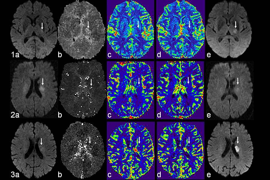
- Förster A, Kerl HU, Wenz H, Brockmann MA, Nölte I, Groden C. Diffusion- and perfusion-weighted imaging in acute lacunar infarction: is there a mismatch? PLoS One. 8:e77428, 2013.
Context Column
Contact

Alex Förster, Md
Working Group Neurovascular Imaging
Department of Neuroradiology
Medical Faculty Mannheim
Heidelberg University
Theodor-Kutzer Ufer 1-3
68167 Mannheim
Phone +49 621 383-2443
alex.foerster@umm.de