
Sie befinden sich hier
Inhalt
Development of multimodal theranostics
Our research objective, which is carried out in cooperation with the Fraunhofer Institute for Manufacturing Engineering and Automation (IPA), Project Group for Automation in Medicine and Biotechnology (PAMB), is the development of theranostics that can be used in vitro and in vivo for the characterization of tumor cells, using multimodal imaging and subsequently for patient-individualized endoradiotherapy. The aim is to carry out work on prostate carcinoma-specific biomarkers, as prostate carcinoma is the most common cancer in men in Germany (26 %) and the third leading cause of cancer death in Germany (10 %).
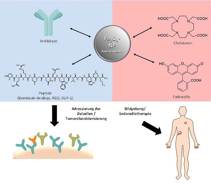
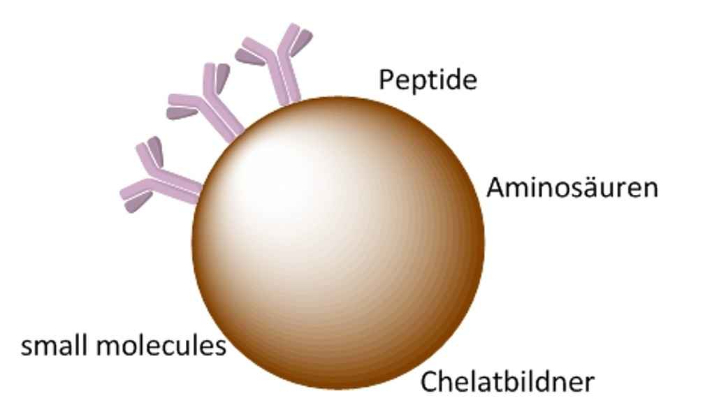
The basis of the theranostics to be developed is the functionalisation of magnetic iron oxide nanoparticles, whereby the functional groups will provide binding possibilities for biomolecules such as antibodies or peptides that specifically bind to receptors (over)expressed on prostate carcinomas by means of specific and efficient reactions. The magnetic iron nanoparticles can be used to conjugate the specific binding ligands for various applications:
- Analysis of receptor expression on tumor cells/target cells. The binding of the iron particle ligands will be quantified after incubation with the cells and thus the target molecule expression on the tumor cell surface will be determined.
- Purification of tumor cells after binding of superparamagnetic iron nanoparticles using magnetic cell separation technologies.
- Use as multimodal contrast agent. The superparamagnetic properties of the iron nanoparticles allow non-invasive and tumor-specific imaging by MRT. Further, by introducing chelators on the surface of the particles, radiolabeling and thus PET imaging as well as use as endoradiotherapeutic agents can be considered. In combination with fluorescent dyes, an additional application in optical imaging is possible.
Functionalized Nanoparticles for Contrast-Enhanced Molecular Imaging with CT
As of yet, for computer tomography (CT), no specifically enriching contrast agents are available, which means the full potential of the method cannot be exercised. Therefore, the aim here is to develop specifically enriching CT contrast agents. For this purpose, for example, certain nanoparticles are suitable which cause a strong weakening of the X-ray radiation and are characterized by high chemical stability, low toxicity and an easily functionalized surface.
Due to these properties, functionalized particles have great potential for use as a basis for molecular imaging using CT. However, for specific enrichment, the particles must be stably coated with different bioactive molecules that specifically accumulate in the target tissue. The contrast media thus obtained are then examined for their suitability in specific CT imaging.
Development of new radiopharmaceuticals for PET diagnostics and therapy of malignant diseases and investigation of the transferability of the results to the human organism.
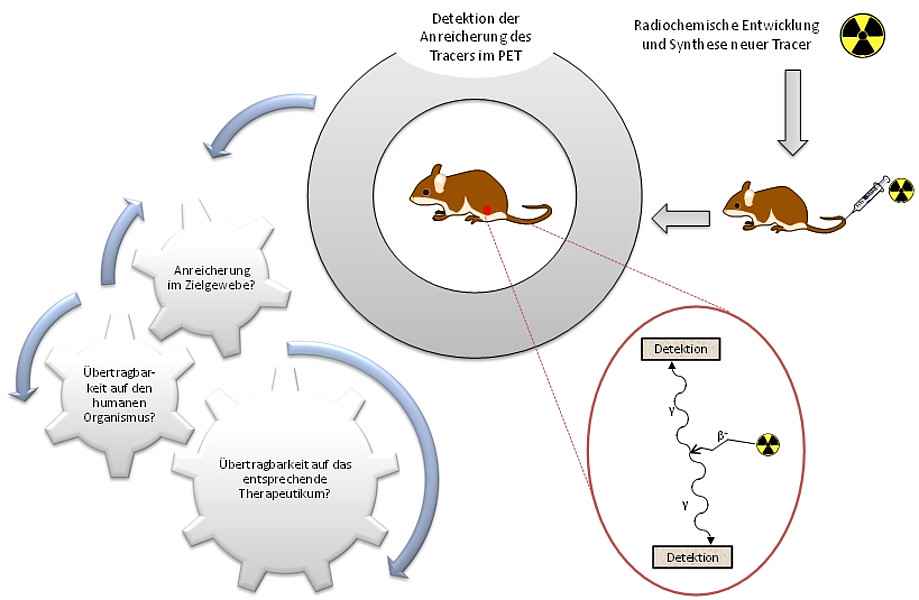
Target-specific molecular imaging using positron emission tomography (PET) has become increasingly important for the diagnosis of various diseases due to its high sensitivity and quantifiability, furthering the growing demand for suitable radiopharmaceuticals. In addition to the development of new positron emitter-labeled radiopharmaceuticals, which allow the visualization of the target structure with high sensitivity and selectivity, another important aspect is the estimation of the distribution of a new radiopharmaceutical in the human organism. This applies not only to PET diagnostics but also to therapeutics for use in endoradiotherapy.
Therefore, here we aim to develop new diagnostic drugs based on different radionuclides for PET imaging of malignant diseases then produce them and examine them in vitro and in vivo. In addition, it is determined by physiologically based pharmacological modeling to what extent the experimental data obtained can be transferred to the human organism, i.e. whether the newly developed radiopharmaceuticals are suitable for human use. This should make it possible to transfer newly developed compounds into clinical application faster and with less risk. Corresponding work will also be carried out with appropriate therapeutic compounds to improve not only the diagnosis but also the treatment of various malignant diseases.
Development of new radiotracers based on 18F-SiFA technology for oncological PET imaging
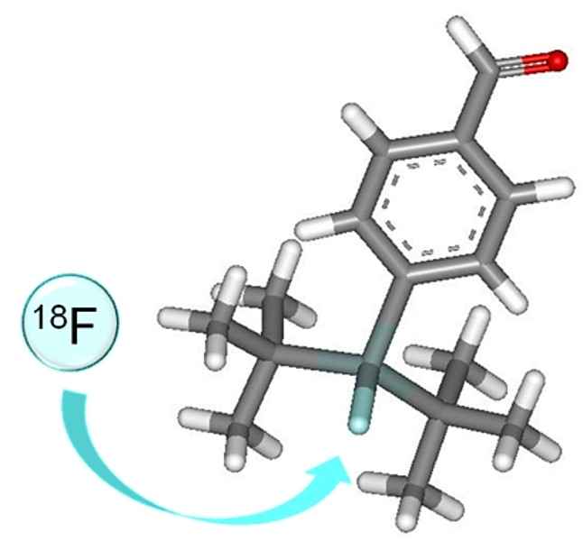
Radioactive labeling reaction, especially bioactive compounds such as peptides with the positron emitter [18F]Fluoride, usually require several reaction and purification steps, whereby the radiochemical syntheses are considered to be partially very complex. With the help of the SiFA component (SiFA = silicon-based fluorine acceptors), which can be introduced directly into peptidic compounds, for example, direct radiolabeling is possible via a single reaction step using an isotope exchange reaction of [19F]fluorine with [18F]fluorine.
So far, this approach has been successfully applied to different tumor affine peptides, and by continuous further development of both the molecule design and the radiolabeling itself, 18F-radiolabeled compounds with partly excellent in vitro and in vivo properties (very good accumulation in the tumor with very low background accumulation) could be obtained. A further advantage of this method is the kit-like, very efficient synthesis of peptide receptor ligands for the personalized diagnosis of oncological problems.
Development of a new 89Zr radiolabeling chemistry for the application of 89Zr in human in vivo PET imaging
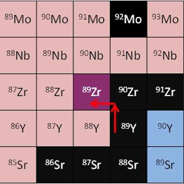
The objective of this research project, carried out in cooperation with Mannheim University of Applied Sciences and Zyklotron AG in Karlsruhe, is the development of new radiotracers based on the radionuclide 89Zr for positron emission tomography.
89Zr represents an advantageous nuclide for PET imaging due to its long half-life of 78 h, its 22.3% positron branching and its mean positron energy, which lies between 18F and 68Ga. Furthermore, it can be obtained at the cyclotron by irradiating stable, high-purity 89Y in an 89Y(p,n)89Zr reaction.
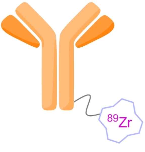
Its half-life makes it particularly suitable for the non-invasive visualization of the biodistribution of slowly accumulating biomolecules, such as antibodies, which offer high tumor specificity and thus achievable high tumor-to-background ratios for the visualization of malignant diseases.
However, as the 89Zr complexation chemistry currently used has disadvantages, especially with regard to the low stability of the 89Zr complexes, this project aims to improve the already known complexing agents in order to optimize the in vivo pharmacokinetics of the 89Zr radiolabeled substances and to exploit the full diagnostic potential of this radionuclide for PET.
Development of new multimeric tumor affine peptides for in vivo detection of malignant diseases using PET
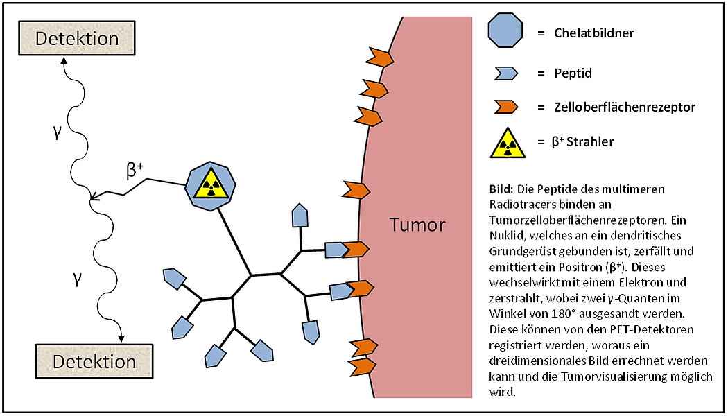
Monomers of receptor affine peptides that specifically bind to receptors overexpressed on tumor cells often show too low in vivo stability or too low tumor-to-background ratios, which limits their use in in vivo PET imaging of malignant diseases.
However, the multimerization of these peptides allows the production of stabilized substances that bind specifically and with high affinity to the tumor and thus allow a high accumulation in the tumor. By radiolabeling with positron radiators, valuable new radiotracers with improved in vivo properties for PET can be obtained.
For this purpose, dendritic basic frameworks are initially produced, on the basis of which the specifically enriching monomeric peptides are multimerized. This is followed by the introduction of a chelating agent and radiolabeling with a positron emitter for in vivo application of the substances. The new peptide multimers thus produced will then be examined in cell experiments for their tumor cell binding properties, whereafter their suitability for in vivo tumor imaging using PET will then be determined.
Apoptosis and its non-invasive PET imaging
This work is to establish an 18F-labeled PET tracer for non-invasive imaging of apoptosis. The non-invasive imaging of apoptotic processes is valuable for clinical treatment, as the severity of the disease and the prognosis for various acute diseases, such as myocardial infarction, can be estimated. Moreover, the therapy of the disease can be monitored and the efficiency of treatment determined. This is particularly advantageous because apoptotic changes usually occur well before morphological changes and the course of a disease can therefore be assessed very early on. Ziel der Arbeiten ist die Etablierung eines 18F-markierten PET-Tracers zur nicht-invasiven Bildgebung von Apoptose. Die nicht-invasive Bildgebung apoptotischer Prozesse ist für die klinische Behandlung wertvoll, da die Schwere des Krankheitsverlaufes und die Prognose bei verschiedenen akuten Erkrankungen, wie beispielsweise Herzinfarkt, abgeschätzt werden kann. Weiterhin kann eine Therapie der Erkrankung verfolgt und die Behandlungseffizienz ermittelt werden. Dies ist vor allem deshalb vorteilhaft, da apoptotische Veränderungen meist deutlich vor einer morphologischen Veränderung auftreten und somit der Verlauf einer Erkrankung sehr frühzeitig abgeschätzt werden kann.
Furthermore, numerous therapy strategies in the treatment of malignant diseases are based on the induction of apoptosis, which makes efficient apoptosis imaging a valuable tool for the representation of the response to therapy.
In vivo apoptosis detection using PET can be achieved with 18F-labeled small molecules that specifically react to changes in apoptotic cells.
Development of 18F-labeled dopamine derivatives for monitoring enrichment in donor kidneys
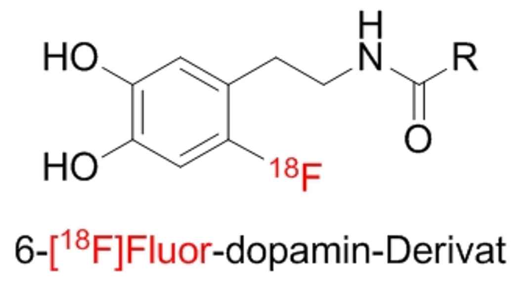
Our objective here is to develop an 18F-labeled, dopamine-based radiotracer for PET imaging.
In clinical operation, kidney donors are treated with dopamine prior to kidney removal for transplantation. Due to the increased level of dopamine in the donor kidneys, significantly fewer dialyses have to be performed in the kidney recipients after transplantation, among other positive effects. However, the duration and mechanism of the protective effect of dopamine treatment are unclear.
Additionally, it has been shown that different derivatives of dopamine have different efficacy with regard to kidney protection and that side effects observed in the case of dopamine can be at least partially avoided.
Therefore, radiotracers based on various dopamine derivatives will be developed and tested for their suitability to elucidate the mechanism of kidney protection and their accumulation and metabolization. Based on the results, targeted substances with even stronger kidney protective effects can then be developed.
