Sie befinden sich hier
Inhalt
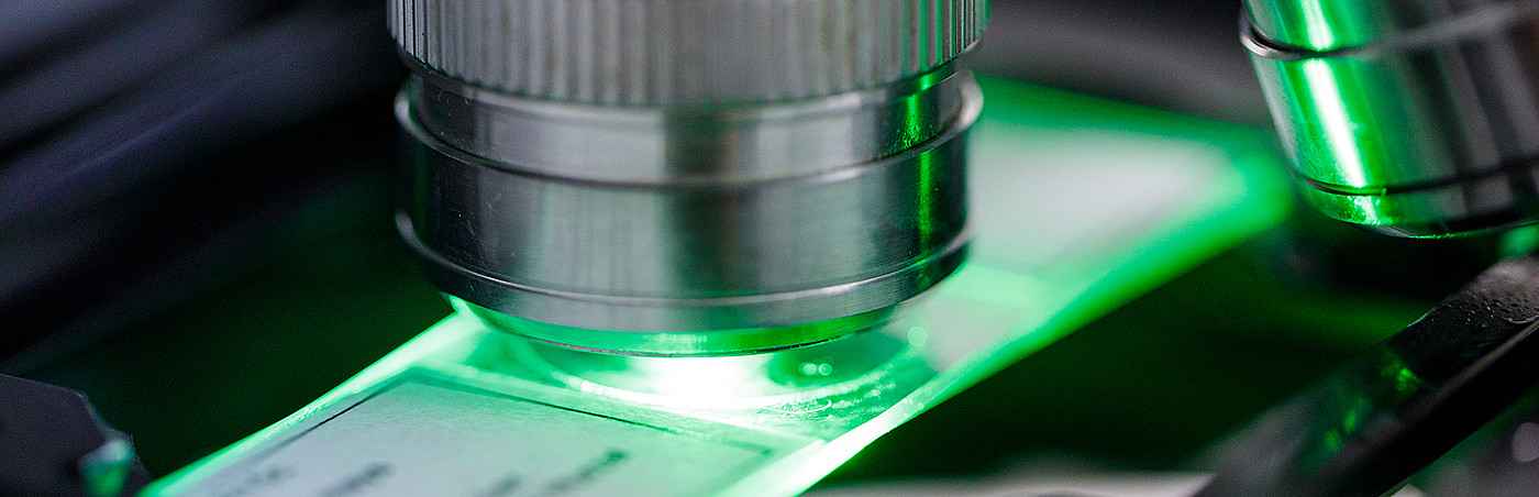
LIMa is a core facility providing advanced microscopy imaging and computational bioimage analysis services to researchers at the Medical Faculty Mannheim of Heidelberg University, as well as to external academic partners.
LIMa integrates state-of-the-art imaging platforms with quantitative and computational image analysis to support modern biological and medical research through the following activities:
- Scientific consultation and user meetings for project planning, technical exchange, and service coordination
- Access to and support for advanced microscopy platforms, including confocal, multiphoton, live-cell, and light-sheet imaging
- Collaborative development and implementation of microscopy imaging methods
- Guidance on quantitative image analysis approaches and established analysis software
- Development of custom image analysis workflows and computational tools in close collaboration with users
- Courses and workshops covering microscopy image analysis, quantitative methods, and scientific programming
Please note that access to LIMa microscopes and devices requires prior service registration.
For more information on computational bioimage analysis activities and methodological development at LIMa, see here.
News
March 2026: Microscopy Image Analysis Course
This course covers key theoretical principles of microscopy image analysis and provides hands-on training with common scientific software such as Fiji. It is designed for PhD and MD students as well as researchers in life and healthcare sciences with no prior experience required. More info…
IEEE ISBI 2025 Best Paper Award
Dr. Qi Gao received the IEEE ISBI 2025 Best Paper Award at the IEEE International Symposium on Biomedical Imaging in April 2025 in Houston, TX, USA, for the paper "Non-Rigid Registration of Time-Lapse Cell Microscopy Images Improved by a Semi-Incremental Optimization Method" (jointly with Karl Rohr). The paper was selected as second best scientific paper out of more than 600 accepted peer-reviewed papers.
Kontextspalte
Core Facility LIMa
Tridomus C410
Ludolf-Krehl-Str. 13-17
68167 Mannheim
Phone +49 621 383-71414
lima@medma.uni-heidelberg.de
Head