Sie befinden sich hier
Inhalt
MicroRNA-induced microangiopathy in diabetes
Supervisor (Mannheim): Hans-Peter Hammes
Co-Supervisor (Groningen): Guido Krenning
Graduate: Chiara Middel
Project description
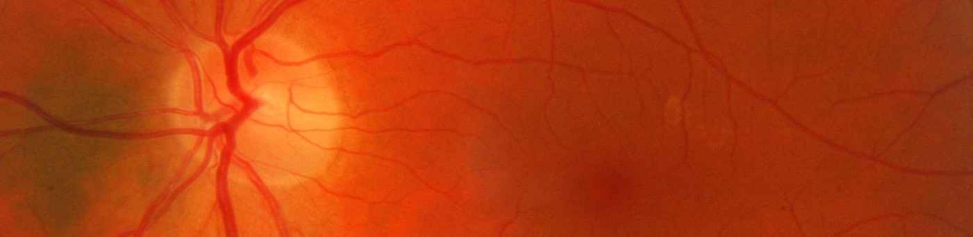
Diabetic retinopathy is a chronic microvascular complication affecting virtually all patients with diabetes. Diabetic retinopathy is characterized by progressive alterations in the retinal microvasculature, i.e. endothelial dysfunction, vascular hyperpermeability, hyperglycemia-induced ROS production, pericyte dropout (Hammes et al. Diabetes 2011. MicroRNAs are small non-coding nucleotides that affect cell function by translational repression of their target genes. MicroRNA dysregulation is associated with diabetic retinopathy (Kovacs et al. Invest. Ophtalmol. Vis. Sci. 2011) and affects endothelial function and angiogenesis (Shantikumar et al. Cardiovasc.Res. 2012). We have recently identified several specific microRNAs that augment endothelial dysfunction by modulating crucial endothelial signaling transduction pathways and hypothesize that dysregulation of microRNAs occurs when diabetic complications develop.
The main aim of this project is to clarify how novel diabetes-related microRNA (diamiR) ‘master switches’ can be manipulated to restore endothelial phenotype and function in diabetic retinopathy. Hereto we aim to (1) identify diamiRs which are associated with diabetic retinopathy and their gene targets, (2) to investigate the influence of diamiRs on the development of endothelial dysfunction and diabetic retinopathy, and (3) to therapeutically modulate diamiR expression to alleviate diabetic retinopathy in a mouse model.
References
- Hammes HP, Feng Y, Pfister F, Brownlee M. Diabetic retinopathy: targeting vasoregression. Diabetes 2011;60:9-16
- Kovacs B, Lumayag S, Cowan C, Xu S. microRNAs in Early Diabetic Retinopathy in Streptozotocin-Induced Diabetic Rats. Invest. Ophtalmol. Vis. Sci. 2011; 52: 4402-9
- Shantikumar S, Caporali A, Emanueli C. Role of microRNAs in diabetes and its cardiovascular complications.Cardiovasc.Res. 2012;93:583-93.
Methods used
Spontaneous and induced animal models of diabetes; laser micro dissection microscopy, Illumina arrays, quantitative retinal morphometry, immunofluorescence and laser scanning microscopy, mass spectrometry, immunoblotting techniques, microRNA in situ hybridization, permeability assays, ROS assays, transfection.
Collaboration Partners
- Guido Krenning, Groningen
- Supervisors of GRK 1874 DIAMICOM in Mannheim, Heidelberg, and Groningen