Sie befinden sich hier
Inhalt

Senior Physician
Professor of Neurological Imaging
Research focus neurological imaging, Department of Neurology
Magnetic Resonance Imaging
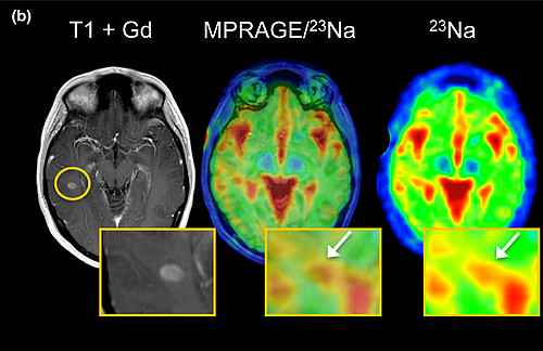
The magnetic resonance imaging group is interested in new magnetic resonance (MR) technologies for the assessment of acute and chronic central nervous system (CNS) disorders with focus on multiple sclerosis, stroke syndromes, subcortical and cortical dementias. In this context the group has applied new methods, such as sodium 23Na MR imaging, developed and performed the MR assessment of the slow diffusion components by Q-space imaging as a more specific approach to detect tissue changes in the normal appearing white matter, applied ASL perfusion imaging and performed non-localised whole brain N-acetylaspartate (NAA) measurements in order to evaluate new pathophysiological aspects of acute and degenerative pathologies.
Central research topics
- Improvement of tissue characterisation by employing quantitative MR imaging methods like magnetisation transfer imaging, diffusion-weighted methods (CHARMED, NODDI), ASL perfusion imaging, and NAA assessments
- Quantitative MR imaging of tissue integrity/myelination status in multiple sclerosis
- Imaging of cortical pathology in multiple sclerosis

Selected research projects
Innovation fund of the Federal Joint Committee: Interactive Web Platform for EmPOWERment in Early Multiple Sclerosis (POWER@MS1) (NCT03968172a). Duration: 2020 – 2023
Baden-Wuerttemberg Ministry of the Economy, Labour and Housing: “Development and integration of a new MR analysis method based on voxel guided morphometry in the evaluation of disease activity in multiple sclerosis” as part of the "AI for SMEs" competition. Duration: 2020 – 2021 (cooperation project with Prof. Lothar Schad, Department of Computer Assisted Clinical Medicine and SMEs)
Biogen GmbH: Non-interventional, multicenter, prospective study investigating an integrated patient management approach in patients with relapsing-remitting multiple sclerosis treated with natalizumab (TRUST). Duration: 2017 – 2021
Selected publications
- Impact of disease-modifying therapies on evolving tissue damage in iron rim multiple sclerosis lesions. Eisele P, Wittayer M, Weber CE, Platten M, Schirmer L, Gass A. Mult Scler. 2022. 1:13524585221106338. doi: 10.1177/13524585221106338
- Characterization of chronic active multiple sclerosis lesions with sodium (23 Na) magnetic resonance imaging-preliminary observations.
Eisele P, Kraemer M, Dabringhaus A, Weber CE, Ebert A, Platten M, Schad LR, Gass A. Eur J Neurol. 2021. 28(7):2392-2395. doi: 10.1111/ene.14873 - Diffusion-weighted MRI in transient global amnesia and its diagnostic implications.
Szabo K, Hoyer C, Caplan L R, Grassl R, Griebe M, Ebert A, Platten M and Gass A. Neurology. 2020. 95(2):e206-e212. doi: 10.1212/WNL.0000000000009783 - Characterization of Contrast-Enhancing and Non-contrast-enhancing Multiple Sclerosis Lesions Using Susceptibility-Weighted Imaging.
Eisele P, Fischer K, Szabo K, Platten M, Gass A. Front Neurol. 2019. 10:1082. doi: 10.3389/fneur.2019.01082 - Temporal evolution of acute multiple sclerosis lesions on serial sodium (23Na) MRI.
Eisele P, Konstandin S, Szabo K, Ebert A, Roßmanith C, Paschke N, Kerschensteiner M, Platten M, Schoenberg SO, Schad LR, Gass A. Mult Scler Relat Disord. 2019. 48-54. doi: 10.1016/j.msard.2019.01.027 - Diffusion-weighted imaging of the dentate nucleus after repeated application of gadolinium-based contrast agents in multiple sclerosis.
Eisele P, Szabo K, Ebert A, Radbruch A, Platten M, Schoenberg SO, Gass A. Magn Reson Imaging. 2019. 58:1-5. doi: 10.1016/j.mri.2019.01.007
Kontextspalte
Contact
Medical Faculty Mannheim
Heidelberg University
Theodor-Kutzer-Ufer 1-3
68167 Mannheim
achim.gass@medma.uni-heidelberg.de
