Sie befinden sich hier
Inhalt
Introduction
By means of magnetic resonance imaging (MRI), descending inhibitory and facilitatory pathways in the spinal cord can be assessed structurally by diffusion MRI (dMRI). Because imaging of the spinal cord is challenging [1] due to small cross-sectional dimensions, sensitivity to physiological motion and bone surrounding the spinal cord, such studies are so far rarely reported [2]. We developed a dMRI phantom of the lumbar spinal cord to optimise dMRI protocols for use in patients with lower back pain and/or paraplegia.
Materials and methods
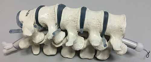
To simulate nerve fibres, a polyamide filament (50 dtex) of 50 μm thickness was selected (Filamentgarn TYPE 611, Trevira GmbH, Bobingen, Germany). The filament was spooled to form a fibre bundle to be placed within a plastic tube. As an emulation of the surrounding spinal segments, a commercially available anatomical model of L1 - L5 (EZ Erler Zimmer, Lauf, Germany) was used. The different fibre phantoms can be easily adjusted and (re)placed into the spinal canal (see Figure 1).
dMRI data acquisition was performed on a 3T MAGNETOM Trio whole-body MR scanner (Siemens Medical Solutions, Erlangen, Germany) using a single-shot spin-echo echo planar imaging sequence with following parameters: TR = 5000 ms, TE = 100 ms, FoV = 210 x 20 mm2, matrix size = 178 x 128, voxel size = 2 x 2 x 2 mm3, no gap, bandwidth = 798 Hz/px. Diffusion weighting was performed in multi-directional diffusion weighting (MDDW) mode along 6 non-collinear directions with b = 1000 s/mm2 and repeated 10 times to enhance signal-to-noise ratio. Additionally, a single non-diffusion weighted volume (b = 0 s/mm2) was acquired at each average.
Results
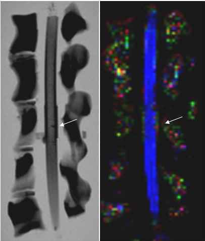
The fibre bundle was cut partially at one side to simulate a spinal cord lesion. The cut is clearly visible at a T1 image of the spinal cord fibre phantom (see Figure 2, left). It is not so clearly visible on a the fractional anisotropy map (see Figure 2, right).
References
[1] Vedantam et al., Neurosurgery, 2014, 74, pp. 1 - 8.
[2] Wang et al., NeuroReport, 2014, 25, pp. 1368 - 1392.
Further reading
-
M. Ruttorf, J. Filip, T. Schaible, M. Weis and F. Zoellner.
White Matter Integrity Differences in 2-year-old Children Treated with ECMO: A Diffusion-Weighted Imaging Study.
Euro J Neurosci, 2025, 61, e70026 -
J. Rausch, M. Ruttorf, L.R. Schad and F.G. Zoellner.
Implementation of a Diffusion Tensor Imaging Phantom of the Lumbar Spinal Cord.
Proceedings of the 32nd ESMRMB, 2015, S465 - S467.