
Sie befinden sich hier
Inhalt
Introduction
Graph theory gives a language for networks. It allows to define networks and to quantify network properties at different levels. In general, networks (or graphs) are represented as sets of nodes N and edges k. The abstract representation of the brain as a graph allows to visualize functional brain networks and describe their non-trivial topological properties in a compact and objective way. Community structure analysis, which detects the groups of regions more densely connected between themselves than expected by chance, is essential for understanding brain network organization and topology [1].
Brain networks demonstrate hierarchical modularity, or multi-scale modularity, i.e. each module contains a set of sub-modules that contains a set of sub-sub-modules etc [2]. We looked into that more closely.
Examples
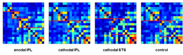
- transcranial direct current stimulation (tDCS) and object recognition
We combined tDCS with a task-related paradigm in fMRI using a repeated measures design [see 3 for further information]. The tool network is organised in a modular way comparable to colour vision which is shown to be automatic, effortless and informationally encapsulated [4]. We treated the tool network as a modular process being a subset of highly functional-connected nodes. Keeping this in mind, we are able to show that tDCS can induce functional brain network reorganisation to the tool network.
Results
Community structure changes depending on stimulation polarity and stimulation site (IPL: inferior parietal lobule, STS: superior temporal sulcus)
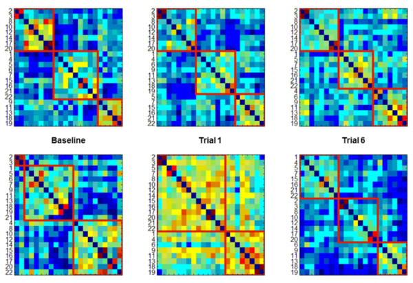
- real-time fMRI in pain processing
We combined real-time fMRI with a painful electrical stimulus to investigate self-regulation of the BOLD response in ten healthy subjects. The subjects were asked to up or downregulate anterior cingulate cortex (ACC) - for training, we used six trials on four consecutive days [see 5 for further information]. There was no strategy given to the subjects beforehand.
Results
Learners (upper row) have ideal community structure right from the start, non-Learners (lower row) do not - they have to acquire it during training (example shown: ACC upregulation)
References
[1] Rubinov & Sporns, NeuroImage, 2010, 52, pp. 1059 - 1069.
[2] Meunier et al., Front Neurosci, 2010, 4, Article 200.
[3] Almeida et al., Cortex, 2017, 94, 176 - 181.
[4] Zeki & Bartels, Phil Trans R Soc B, 1998, 353, pp. 1911 - 1914.
[5] Rance et al., Hum Brain Mapp, 2014, 35, 5784 - 5798.
Further reading
- M. Ruttorf, Z. Tal, L. Amaral, F. Fang, Y. Bi and J. Almeida. Neuroplastic changes in functional wiring in sensory cortices of the congenitally deaf: A network analysis. Human Brain Mapp 2023, 44, p 6523-6536.
- M. Ruttorf, S. Kristensen, L.R. Schad and J. Almeida. Transcranial Direct Current Stimulation Alters Functional Network Structure in Humans - A Graph Theoretical Analysis. IEEE T Med Imaging, 2019, 38, 2829 - 2837.
- M. Ruttorf, S. Kristensen, L.R. Schad and J. Almeida. Transcranial direct current stimulation alters functional network structure in humans. 2018, bioRxiv 296657, doi: 10.1101/296657.
- M. Rance, M. Ruttorf, F. Nees, L.R. Schad and H. Flor. Real time fMRI feedback of the anterior cingulate and posterior insular cortex in the processing of pain. Hum Brain Mapp, 2014, 35, 5784-5798.
- M. Bucolo, M. Rance, A. Muscarello, A. Spampinato, M. Ruttorf and H. Flor. Functional Neuro-Imaging and Brain Networks. International Journal of Bioelectromagnetism, 2012, 14, 100 - 107.