Sie befinden sich hier
Inhalt
The group performs research on new methods of image formation in MRI, Computer Tomography (CT) and X-Ray imaging, especially in physiological / functional imaging of the human body as well in preclinical projects. Furthermore, we research on methods to automatically analyse the acquired data by means of medical image processing and pattern recognition using machine learning techniques to quantify the measured physiological parameters.
We also perform research related to image guided interventions.
Recent projects
MOLECULAR INNOVATIVE IMAGING FOR INDIVIDUALIZED DIAGNOSTICS
The clear identification and characterization of therapy-relevant focal findings using innovative diagnostic methods forms the basis for minimally invasive therapy (including radionuclide therapy) for patients with oligometastases in the closed-loop process of M²OLIE. It also allows a prediction of the expected therapy response and the early detection of patients who do not respond to a specific therapy (non-responders). The aim of the joint project M²IBID is to explore these basic prerequisites for determining an effective therapy in the sense of “precision medicine”. The techniques of diagnostic and interventional imaging and molecular mass spectrometry (MS) bioanalytics will be integrated more deeply and clinically evaluated. The innovative research goal is to ideally combine all this information to explore novel tumor characterizations using radiomics approaches and machine learning techniques that improve patient-specific intervention.
Persons involved: Frank G. Zöllner (PI), Dominik Bauer, M.Sc., Tom Russ, M.Sc., Christian Toennes, M.Sc.
Funding: German Federal Ministry of Education and Research (BMBF)
MAGNETIC RESONANCE IMAGING BIOMARKERS FOR KIDNEY DISEASE - Coordinating research on renal MRI biomarkers for clinical practice, drug development and basic research.
The rising prevalence of Chronic Kidney Disease (CKD) poses a major public health challenge affecting >10% of the population. There exists an urgent need for better biomarkers to identify patients that are at risk of progression, or are likely to respond to (candidate) therapeutics. Magnetic Resonance Imaging (MRI) biomarkers can help to fill this gap, as they are uniquely able to track disease progression and treatment effects in the tissue itself and in a non-invasive manner.
Persons involved: Frank G. Zöllner (WP2 lead)
Renal MRI standardisation to improve CKD management
Persons involved: Frank G. Zöllner (PI), Aish Raj, M.Sc. , Anika Strittmatter, M.Sc.
Funding: German Federal Ministry of Education and Research (BMBF) under the frame of ERAPerMed
Prediction of treatment response and outcome in locally advanced rectal cancer using radiomics and deep learning: an example case to demonstrate a general purpose deep-learning-based processing pipeline for image classification
In the last decade, rectal cancer has become the third leading cause of cancer death. Although diagnostic and treatment options have improved in recent years, rectal cancer remains a highly heterogeneous disease in terms of treatment response and outcome.To date, only clinical and magnetic resonance imaging (MRI) criteria have been used to make treatment decisions once the diagnosis is confirmed. Although MRI has become the standard diagnostic tool for local staging of rectal cancer, it does not provide information on intratumor heterogeneity or molecular subtypes. Consequently, novel imaging biomarkers are urgently needed to better characterize subtypes of rectal cancer and to better predict treatment response.Texture analysis, radiomics, and deep learning strategies are increasingly being used to address these challenges and improve patient care. However, patient cohorts have been relatively small in many previous studies, often lacking a validation cohort, and imaging data have been collected at a single institution or at different centers with similar MRI scanners, making it impossible to assess the generalizability of the trained models. Modern radiomics techniques are therefore increasingly shifting to newer developments in deep learning.The aim of this study is to develop a radiomics- and deep learning-based signature for rectal cancer capable of decoding different tumor phenotypes and predicting therapeutic response in a non-invasive manner in the context of histopathology and genomics/clinomics
Persons involved: Frank G. Zöllner (PI), Steffen Albert, M.Sc.
Funding: German Research Foundation (DFG)
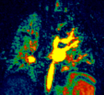
Quantitative pulmonary function testing in 2-year-old children after congenital diaphragmatic hernia using Fourier decomposition and magnetic resonance imaging
Congenital diaphragmatic hernia (CDH) results in displacement of abdominal organs into the thoracic cavity and leads to underdevelopment of the lungs, followed by pulmonary hypoplasia. Pulmonary hypoplasia and pulmonary hypertension are the main factors in the survival risk of the child with CDH. Pulmonary hypoplasia not only affects the formation air vessels but also leads to dysfunction of the capillary bed. In healthy children, the number of aveoli grows until the age of 3 years. Therefore, observation of lung development in children after correction of CDH is important to assess long-term effects for the child. The extent to which the lung can regenerate after CDH is currently unknown. However, lung perfusion (Q) and ventilation (V) can be measured noninvasively on MRI without the use of contrast agents by means of the so-called Fourier decomposition (FD). Our own preliminary work has shown that quantitative measurement of V/Q is possible in adults. The aim of this proposal is to implement this technique for 2-year-old children and to evaluate the clinical added value. In a first step 2D and in a second step 3D imaging with appropriate postprocessing will be developed, both at 1.5T and 3.0T. From a clinical point of view, the diagnostic quality of the images will be determined, whether they can detect differences in perfusion and ventilation with respect to pulmonary hypolasia, and ultimately the significance of this procedure for the follow-up of children will be determined.
Persons involved: Frank G. Zöllner (PI), Efe Illicak, M.Sc.
Funding: German Research Foundation (DFG)
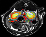
Characterization of hepatocellular carcinoma heterogeneity by radiomics - A systematic test in the transgenic mouse model.
The integrative assessment of molecular and genetic data together with multiparametric image data sets lays the foundation for a completely novel diagnostic approach that will not only allow improved disease characterization but also outcome prediction. This approach thus creates the basis for individualized therapy approaches in the sense of precision medicine. Despite the high expectations for radiomics, the evaluation is currently still in a trial phase with many factors influencing the analyses that are not yet understood.hepatocellular carcinoma is a very inhomogeneous disease whose diagnosis to date is subject to known limitations, both in the primary situation and under therapy. Clear imaging characteristics that allow the detection of intratumoral heterogeneity as well as a clear assessment of treatment response do not exist to date. The aim of the present application is to perform a systematic radiomic analysis to define imaging and non-imaging biomarkers that identify treatment responders and enable outcome prediction.
Persons involved: Frank G. Zöllner (PI), Simon Reichert, M.Sc.
Funding: German Research Foundation (DFG)
Recent publications
-
A. Adlung, N. Paschke, A. Golla, D. Bauer, S. Mohamed, M. Samartzi, M. Fatar, E. Neumaier-Probst, F. Zöllner and L. Schad.
23Na MRI in ischemic stroke: Acquisition time reduction using postprocessing with convolutional neural networks.
NMR Biomed, 34 (4), p.e4474 2021. -
D. Bauer, T. Russ, B. Waldkirch, C. Tönnes, W. Segars, L. Schad, F. Zöllner and A. Golla.
Generation of annotated multimodal ground truth datasets for abdominal medical image registration.
Int J Comput Ass Rad, 16 (8), pp.1277-1285 2021. -
D. Bauer, A. Adlung, I. Brumer, A. Golla, T. Russ, E. Oelschlegel, F. Tollens, S. Clausen, P. Aumüller, L. Schad, D. Nörenberg and F. Zöllner.
An Anthropomorphic Pelvis Phantom for MR-guided Prostate Interventions.
Magn Reson Med, , p.in Press 2021. -
S. Bech, H. Qi, C. Mariager, E. Hansen, E. Ilicak, F. Zöllner and C. Laustsen.
The number of glomeruli and pyruvate metabolism is notstrongly coupled in the healthy rat kidney.
Magn Reson Med, , p.in Press 2021. -
A. de Boer, G. Villa, O. Bane, M. Bock, E. Cox, I. Dekkers, P. Eckerbom, M. Fernández-Seara, S. Francis, B. Haddock, M. Hall, P. Barrientos, I. Hermann, P. Hockings, H. Lamb, C. Laustsen, R. Lim, D. Morris, S. Ringgaard, S. Serai, K. Sharma, S. Sourbron, Y. Takehara, A. Wentland, M. Wolf, F. Zöllner, F. Nery and A. Caroli.
Consensus-based technical recommendations for clinical translation of renal phase contrast MRI.
J Magn Reson Imaging, , p.in Press 2021. -
V. Groß, K. Zahn, K. Maurer, L. Wessel, T. Schaible, S. Schoenberg, C. Weiß, F. Zöllner and M. Weis.
MR lung perfusion measurements in adolescents after congenital diaphragmatic hernia: correlation with spirometric lung function tests.
Eur Radiol, p.in Press 2021.
Kontextspalte
Head of Group


House 3, Level 4, Room 112
Phone +49 621 383-5117
Fax +49 621 383-5123
frank.zoellner@medma.uni-heidelberg.de