Sie befinden sich hier
Inhalt
J. Zapp1, S. Schmitter2, L. R. Schad1
Introduction
Acoustic scanner noise during image acquisition is a well-known problem in functional MRI (fMRI). The noise is created by gradient coil switching and causes an unwanted BOLD signal in the auditory cortex [1]. Most of the time, echo-planar imaging (EPI) is used for fMRI. However, this sequence produces high acoustic noise due to fast switching of trapezoidal gradients. The sound pressure level (SPL) of such EPI sequences on a 3T-scanner can reach up to 130 dB [2]. This effects fMRI at high resolution seriously. High resolution in EPI can be achieved by a parallel acquisition technique (PAT).
The aim of this work is to extend an existing silent sinusoidal EPI (sEPI) sequence by means of PAT. This technique allows for doubling the current resolution (64 x 64 matrix) of the sEPI at approximately the same acquisition time. The developed sequence is evaluated in an auditory fMRI experiment. The combination of the sEPI sequence with PAT results in a high resolution sequence that reduces the SPL compared to the conventional EPI sequence with PAT.
Materials and methods
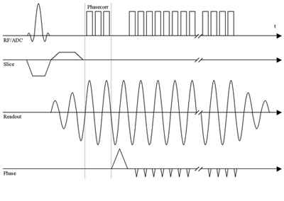
In this work a sEPI sequence is used that replaces the trapezoidal readout gradient of EPI by a sinusoidal readout gradient which is depicted in Fig. 1. The sEPI shows to be an effective approach to reduce the SPL of the scanner while maintaining the same imaging parameters like an EPI sequence [3].
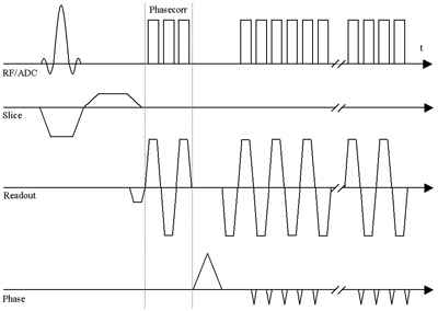
In order to allow for silent imaging with high resolution and a short echo time, the sEPI is extended by a parallel acquisition technique (PAT). Here, GRAPPA [4] is used as PAT. PAT allows for reducing acquisition time, since the k-space is undersampled. The gained time is used to increase the resolution while keeping the echo time constant. The missing information due to undersampling is obtained by a PAT reference scan (see Fig. 2). The reference scan is implemented as a single-shot EPI with trapezoidal readout gradients. Rising noise there from does not effect the actual image acquisition, since the reference scan is executed separately, right before the undersampling sEPI acquisition.
The sEPI PAT sequence and the corresponding image reconstruction for regridding the sinusoidal sampling is implemented on a 3T-scanner (Magnetom Tim Trio, Siemens Medical Solutions, Germany). A 12-channel head matrix is used as receiver coil.
The SPL measurements are performed by means of an optical microphone (MO 2000, Sennheiser electronic GmbH & Co. KG, Germany) mounted inside the receiver coil.
In order to assess the silent sEPI sequence for high resolution fMRI, a first pilot experiment is conducted on a healthy volunteer. This experiment uses an external stimulus with a tonal frequency of 1768 Hz which matches a strong frequency component in the noise spectrum of a conventional EPI sequence using a bandwidth of 1396 Hz/Px and PAT. The aim is to find out whether the sEPI PAT sequence can achieve a higher significance of activation by the stimulation than the conventional EPI with PAT. A simple block design is used including eight blocks during the presentation of the stimulus and nine blocks at rest, beginning with a rest block. Data analysis is performed using SPM5 (Welcome Department of Cognitive Neurology, Institute of Neurology, University College London, London, UK) with motion correction (realignment), spatial smoothing (Gaussian-Window, FWHM = 5 mm), normalization to a standard EPI template (MNI template) and a cluster size of 10 voxels.
Results
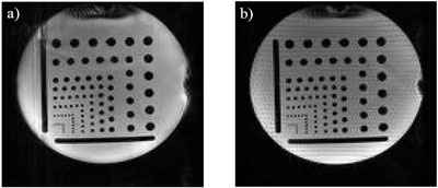
Fig. 3 shows that the high resolution image of the developed sEPI PAT sequence is comparable to the EPI PAT sequence. However, measurements of the average SPL result in a lower SPL for the sEPI PAT sequence. The minimum average SPL for varying bandwidths is achieved when using the imaging parameters matrix: 128 x 128, FOV: 220 x 220 mm², TE: 39 ms, TR: 74 ms, BW: 1396 Hz/Px for both sequences and yields a SPL of (78.2 ± 1.3) dB for sEPI PAT and (88 ± 6) dB for EPI PAT. This means a reduction by 10 dB. Since the SPL is proportional to 20 log p with p being the sound pressure, the sound pressure is reduced by a factor of three when using sEPI PAT instead of EPI PAT.
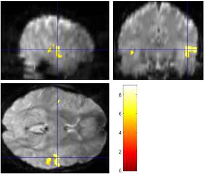
The fMRI experiment gives higher activation for the sEPI PAT with tmax = 8.9 than for the EPI PAT (tmax = 5.1). When the level of significance is set to p < 0.05 (FWE corrected) (threshold tth = 5.2), significant activation is found for the sEPI PAT (see Fig. 4), but no activation at all for the EPI PAT sequence.
Discussion
The new sEPI PAT sequence has comparable imaging parameters as the conventional EPI PAT. The important advantage of the sEPI is the reduction of SPL by 10 dB.
A first pilot fMRI experiment showed a significant higher level of activation of the auditory cortex for the sEPI PAT sequence than for the EPI PAT sequence. However, further experiments have to be conducted to get more meaningful statistics.
1 Computer Assisted Clinical Medicine, Faculty of Medicine Mannheim, University of Heidelberg, Mannheim, Germany,
2 Medical Physics in Radiology, German Cancer Research Center, Heidelberg, Germany
References
[1] Bandettini et al. [1998] Magn. Reson. Med. 39(3):410-416;
[2] Foster et al. [2000] J. Magn. Reson. Imaging 12(1):157-163;
[3] Schmitter et al. [2008] MAGMA 21(5):317-325;
[4] Griswold et al. [2002] Magn. Reson. Med. 47(6):1202-1210.