Sie befinden sich hier
Inhalt
E. Wiener, L. R. Schad, K. Baudendistel, E. Müller*, W. J. Lorenz.
Introduction
Functional magnetic resonance imaging at high temporal resolution is a useful method for tracing stimulus induced MR-signal changes. With a conventional FLASH imaging techniques time resolution is limited to 5-10 s. EPI techniques allow increased temporal resolution, but suffer from inter-scan delays. We have developed a new method to improve the temporal resolution up to 80 ms without reducing spatial resolution [1,2], using a modified FLASH technique. Like in ECG-gated acquisitions, data-sampling and stimulus presentation is repeated until all phase encoding steps are collected. Event-related fMRI could be performed with a temporal resolution of 80 ms for a serie of 128 sequential images or with a temporal resolution of 320 ms by simultaneously measuring four slices with a serie of 32 sequential images.
Materials and methods
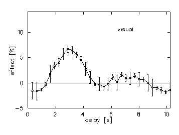
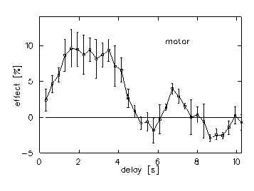
MR images of the visual and motor cortex are obtained at 1.5 T (MAGNETOM 63/84 SP, Siemens). The measuring sequence is a modified 3D, rf-spoiled and first order flow compensated FLASH-sequence (head coil, 4 slices, 4 mm slice-thickness, TR/TE/? /matrix = 320 ms/ 60 ms/ 40°/, 64 ? 128). Total scantime is 10 min 55 s. In this modified sequence the 3D gradient table was removed and the 3D partition-loop acts as a time-loop for the sequential measurement of 128 or 32 consecutive images at the same slice position. We used a TTL-signal coming from the gradient board at the beginning of every line-loop and a self-made ‘TTL-Divider-Box’ for the event triggering. After every 128th TTL-signal a single white light flash (5 ? s), seen by the volunteer in the dark scanner room or an acoustic signal for a single finger to thumb movement of the left hand is given. The MR-signal response can be observed over 32 ? 320 ms or 80 ? 128 ms = 10.24 s. Event-related fMRI examinations were carried out on 10 healthy right handed volunteers (five visual, five motor). Each stimulation experiment covered four contiguous slices, resulting in a total cross section of 16 mm. Activated regions were calculated on pixel-by-pixel basis by correlation of the MR-signal time course with a theoretical response function (time-shifted box-car function). Accidental correlation was minimized by thresholding ( cc > 0.65, ? = 0.0002). Based on correlation coefficient (cc)-maps regions of interest were defined in each subject and the percent signal change was calculated. Averaged signal time courses (? SD) of the five visual and five motor stimulation experiments with significant increased MR-signal responses are presented (Fig. 2 ,3).
Results
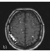
Significant activation occurred predominantly in areas occupied by the gray matter of visual cortex (Fig. 1a), primary right motor and sensory cortex (Fig. 1b). The averaged response magnitudes is about 6% after visual stimulation and about 10% after motor stimulation. While visual response is delayed by about 2 s, motor response shows an instantaneous increase of the MR-signal. Responses are decayed after about 5 s in both cases (Fig. 2, 3).
Conclusions
Event-related fMRI combines the advantages of conventional FLASH-sequences like high spatial resolution and good signal to noise ratio with high temporal resolution. Our data demonstrate that this technique is an interesting tool for resolving some temporal features of cerebral activation.
References
[1] Schad, L.R., Wiener, E., Baudendistel, K., Müller, E., Lorenz, W.J., Magn. Reson. Imag. 6, 899 (1995).
[2] Wiener, E., Schad, L.R., Baudendistel, K., Essig, M., Müller, E., Lorenz, W.J., Magn. Reson. Imag. (submitted) (1995).