Sie befinden sich hier
Inhalt
P. M. Heiler1, S. Konstandin1, L. R. Schad1
Introduction
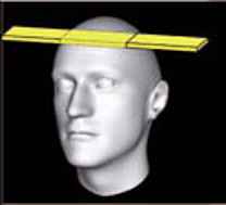
Multi-Echo FLASH imaging in combination with echo train shifting [1,2] offers BOLD sensitivity for matrix sizes up to 256 x 256 leading to in-plane resolutions of about 1 mm2. Long measurement times and low Signal-to-Noise-Ratio (SNR) preclude BOLDfMRI going to higher resolutions. Hence, only a reduction in the Field-of-View (FOV) further increases spatial resolution. Latter, however, is limited by aliasing artifacts due to signals from outside of the FOV. 2D-selective excitation [3] in slice- and phase encoding direction, as shown in Fig. 1, allows for small inner FOV sizes resulting in increased spatial resolution. Recently, applications with two-dimensional pulse profiles such as spiral [4] or blipped planar [5] excitation trajectories were reported. In this study, latter one was chosen because the limiting side excitations only occur in one single dimension.
Methods
The design of the 2D-selective excitation pulse was based on a duration of 27.5 ms, spatial dimensions of 50 mm in read direction and 4 mm in slice direction and a flip angle of 50°. kmax,x was set to 2.5 cm-1, kmax,y to 0.84 cm-1 and Dky to 0.04 cm-1.
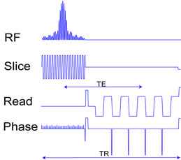
For data acquisition, the Double Contrast Echo Train Shifted Multi-Echo FLASH sequence [2] was chosen with the following sequence parameters: TE=38 ms, TR=126 ms. The matrix size was 192 x 114 leading to an in-plane resolution of 0.5 x 0.5 mm2.
For data acquisition, the Double Contrast Echo Train Shifted Multi-Echo FLASH sequence [2] was performed on a 3 T (Trio, Siemens, Erlangen, Germany) using a matrix head coil with the following sequence parameters: TE=38 ms, TR=126 ms, flip angle α=50°. The matrix size was set to 192 x 114 leading to an in-plane resolution of 0.5 x 0.5 mm2. To demonstrate BOLD-sensitivity, an initial BOLD-experiment was performed by acquiring 200 images in 2.9 s/image. Functional activation was achieved by a visual stimulation task showing a checkerboard alternating with a gray screen (5 images in 14.5 s each).
Functional results were processed and analyzed with Statistical Parametric Mapping 5 (SPM5). The images were realigned to the average image (Fig. 3) and subsequently smoothed with a 2 mm Gaussian Kernel. The alpha error was set to α=0.01 and minimal shown cluster size was k=10 pixels.
Results
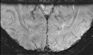
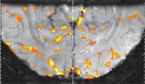
Fig. 3 shows the average image of 200 acquisitions during the fMRI experiment. It should be noted that the image is free of noticeable artifacts at an in-plane resolution of 0.5 x 0.5 mm2. Activation maps of the human brain (Fig. 4) show BOLD-signal only close to the venules.
Discussion
We presented a high resolution T2*-weighted acquisition sequence in combination with a two-dimensional excitation pulse suitable for functional experiments. Its feasibility includes both, high resolution morphological T2*-weighted imaging with high contrast to the venule system (Fig. 3) and local high resolution functional studies (Fig. 4). Latter showed activation only in areas close to venules instead of a blurred activation-signal in the whole cortex, as achieved with generic EPI-BOLD sequences.
1 Computer Assisted Clinical Medicine, University of Heidelberg, Mannheim, Germany
References
[1] Voit D, Frahm J. NMR Biomed 18, 481-488 (2005)
[2] Heiler P et al, Proceedings of the ISMRM 15, 1940 (2007)
[3] Haase A et al, J. Magn Reson 67, 258-266 (1986)
[4] Pauly J et al, J. Magn Reson 81, 43-56 (1989)
[5] Rieseberg S et al, Magn. Reson. Med. 47, 1186-1193 (2002)