Sie befinden sich hier
Inhalt
Functional Magnetic Resonance Imaging of Sensorimotor Cortex at 1.5T: A Comparison of Functional Venography and BOLD-Imaging
K. T. Baudendistel, J. R. Reichenbach, R. Metzner, J. Schröder, L.R. Schad
Introduction
The measurement of brain activation using blood oxygen level dependent contrast (BOLD, 1) relies on the signal change arising from activation-induced decrease in paramagnetic deoxyhemoglobin concentration. This change is not limited to the activated region's parenchyma, but extends towards offleading venous vessels. Imaging of venous vessels at high spatial resolution can be achieved, using the deoxyhemoglobin concentration as source of image contrast (2). Venograms obtained in rest and during a finger tapping paradigm reflect changes in vessel contrast near the regions delineated by brain activation maps obtained with conventional BOLD-EPI-fMRI.
Methods
Mapping of venous structure was performed using a 1.5T clinical EPI-whole body scanner (Magnetom Vision, Siemens Medical Systems, Germany) equipped with the standard head coil. The 3D-gradient-echo sequence described in (2) was used for MR-venography with FOV=240mm, 16 3D-partitions (effective slice thickness=2mm), NEX=2, TA=10min, and a matrix size of 512x320. For improvement of SNR, minimum intensity projections of three consecutive slices were calculated, resulting in 6mm thick slices. A venogram was acquired in resting state followed by a second, obtained during performance of self-paced finger tapping of one hand. BOLD-EPI-fMRI was performed using a FID-echo planar imaging (EPI) sequence with same FOV, TH=3mm, MAT=128², TE=54ms. Twelve consecutive slices were acquired every 3s. fMRI series of 60 images were acquired in cycles of ten images in resting state followed by ten images during task performance. The activation paradigm consisted of the finger tapping described above. Prior to brain map calculation, a motion correction was performed using the MCW-AFNI software package (3). Brain activation maps were calculated using an unpaired Student's t-test. To obtain comparable slice thickness, two consecutive t-maps were averaged.
Results
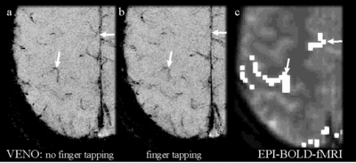
Four healthy volunteers (age 20-30ys) were investigated. All volunteers showed a reduction in venous vessels contrast near the regions of brain activation. Finger tapping resulted in an increased activation of the corresponding sensorimotor cortex and the supplementary motor area (see arrows). A Student's t brain activation map superimposed on the anatomy is presented by Fig.1c. Only pixels exceeding Student's t of 2.0 are shown. The venograms show a corresponding reduction in venous vessel contrast, corresponding to a decrease in the concentration of deoxyhemoglobin under task performance (Fig. 1b) as compared to the resting condition (Fig. 1a).
Conclusions
MR venography allows direct observation of deoxygenation concentration change under task performance with high spatial resolution. Combination of venography and BOLD-fMRI-experiments may offer possibilities for separation of venous vessel and brain parenchyma contribution to the observed BOLD-signal, leading to improved assessment of brain tissue activation.
References
[1] Ogawa, S., Lee, T.M., Kay, A.R., Tank, D.W. Proc. Natl. Acad. Sci. USA 1990, 87: 9868-9872
[2] Reichenbach, J.R., Venkatesan, R., Schillinger, D.J., Kido, D.K., Haacke, E.M. Radiology 1997, 204:272-277
[3] Cox, R.W. Computers and Biomedical Research, 1996, 29:162-173