3D Contrast Enhanced Renal MR Angiography
Multiphase 3D Contrast Enhanced Renal MR Angiography: A Comparison between Cartesian and Spiral Readout
Multiphase 3D Contrast Enhanced Renal MR Angiography: A Comparison between Cartesian and Spiral Readout
M. Amann, F. Floemer, M. Bock, S. O. Schoenberg, L. R. Schad
3D Contrast enhanced Magnetic Resonance Angiography (3D CE-MRA) offers an adequate morphological representation of the renal vasculature in a minimal invasive way [1]. To obtain pure arterio- and venograms, multiphase angiography has been established [2]. The short acquisition time per single phase leads to lower spatial resolution and anatomic coverage.
Spiral MRI with its efficient sampling of k-space is a promising technique to increase resolution without increasing acquistion time. Additionally, spiral MRI is less sensitive to flow artifacts due to the attainable short echo times and the intrinsic compensation of higher gradient moments [3].
The purpose of this work was to compare a standard multi-phase 3D CE-MRA technique [4] with a sequence using inplane spiral readout with respect to the attainable resolution at comparable acquistion time.
The multiphase 3D CE-MRA of the renal arteries were carried out in seven volunteers with a 1.5 T whole body scanner (Magnetom Vision, Siemens, Germany). For each volunteer a contrast bolus of 0.2 mmol/kg bodyweight was administred (Magnevist?) by automatic infu-sion (Tomojet CAI, Doltron). All measurements were performed with a phased array torso coil.
With the spiral MR sequence a conventional phase-encoding gradient in partition direction was combined with an in-plane spiral readout [5]. The spiral trajectories were slew rate optimized with respect to the chosen number of interleaves and to the selected field-of-view (FOV) [6]. Per partition, 16 interleaved spirals with a duration of 15.36 ms were used. For each spiral, 2048 data points were sampled. The other parameters were: TR/ TE/ a = 19.0 ms/ 1.3 ms/ 60°. A postprocessing deblurring algorithm was used for the spiral raw data to correct the effects of B0 inhomogeneities [7].
The conventional MRA sequence was a 3D FLASH with TR/ TE/ a = 3.2 ms/ 1.2 ms/ 30° and asymmetric k-space sampling in readout- and phase-encoding direction. In both sequences the same asymmetric k-space sampling in partition direction was used. For a 80 mm coronal slab with 44 interpolated partions an acquistion time of 6 s (spiral MRI) or 7 s (FLASH) were obtained for a single phase. Five phases were acquired subsequently. With the 3D FLASH a nominal resolution of 1.94 x 1.37 mm2 was achieved at a field-of-view (FOV) of 260x350 mm2. With the spiral technique, an isotropic reso-lu-tion of 1.39 x 1.39 mm2 at a FOV of 350x350 mm2 was attained.
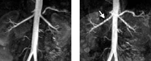
Despite shorter acquisition time, the spiral 3D CE-MRA attains higher resolution than the standard technique. Fig. 1 shows the maximum intensity projection (MIP) at the arterial phase. The FLASH arteriogram (top) shows step artifacts (white arrow) caused by the reduced resolution in phase-encoding direction (left to right).
In this study we demonstrated the efficiency of spiral read-out for multiphase 3D CE-MRA. In spite of the higher effort for reconstruction and correction of the raw data, the spiral technique is a promising tool for further reduction of acquisition time without losing resolution. The focus of future work will now be the application of spiral 3D CE-MRA to pathological processes.
Abt. Biophysik und Medizinische Strahlenphysik, Deutsches Krebsforschungszentrum (dkfz), Heidelberg,Germany
[1] Prince M. R., JMRI 8(3), 511-516, 1998
[2] Schoenberg S. O. et al., JMRI, 10(3), 314-316, 1999
[3] Van Tyen R., Saloner D., 6th ISMRM, 779, 1998
[4] Schoenberg S. O. et al., Radiology 211(3), 667-679, 1999
[5] Amann M., Bock M., Schad L. R., 7th ISMRM, 158, 1999
[6] King K., Foo T., Crawford C., MRM 34, 156-160, 1995
[7] Noll D. C. et al., MRM 25, 319-333, 1992