Sie befinden sich hier
Inhalt
M. Bock, S. O. Schönberg, G. Laub1, M. V. Knopp, L. R. Schad
Introduction
MR CINE phase contrast flow measurements in the renal arteries can be performed in a breathhold with high temporal and spatial resolution. High spatial resolution is required, because the renal arteries have diameters of several millimeters only. Sufficient temporal resolution is necessary, because stenotic flow reductions manifest in subtle changes of the flow profile [1]. Especially the presence of the early systolic peak (ESP), a characteristic feature in the velocity-time curve of about 50 ms duration, is related to the grade of stenosis.
With segmented echo-planar (SEPI) sequences both resolution requirements can be fulfilled [2]. Additionally, acquisition times can be kept as short as a breathhold, so that measurement errors due to renal artery respiratory motion are minimized. Unfortunately, segmented EPI sequences use very strong gradients, that induce high voltages in conventional ECG leads. Therefore, a dead-time of about 50-100 ms has to be waited after the last gradient switching to reliably detect an R-wave in the ECG signal. This results in an incomplete measurement of the flow pattern during the cardiac cycle.
In this pilot-study, an optically decoupled ECG-system [3] was used, that is less susceptible to induced currents, and which allows to monitor the ECG signal even during EPI data acquisition. With this ECG-system, a conventional FLASH CINE phase contrast sequence was compared with a SEPI sequence with prospective and retrospective gating in the evaluation of renal artery blood flow.
Materials and methods
The SEPI phase contrast sequence was implemented on a 1.5 T whole body scanner (SIEMENS Vision, Erlangen, Germany) using the integrated body coil for rf-excitation and a 4-element phased array coil for signal reception. 16 alternating gradient echos were acquired per rf-excitation using blipped gradients for phase encoding. Within each echo-train k-space trajectories started at the center of k-space going to either high positive or negative k-space frequencies. At a readout bandwidth of 977 Hz/pixel, an echo time for the first echo of 3.8 ms and an echo spacing of 1.6 ms a repetition time TR of 31 ms was achieved using a resonant gradient system (n = 833 Hz).
Before image reconstruction, a linear phase correction was performed on each line in k-space with data from 2 non phase-encoded pre-scans. The sequence was sensitized to flow perpendicular to the imaging plane with a VENC of ±74 cm/s.
With prospective gating, flow compensated and flow sensitized scans were acquired in alternate RR-intervals. At a matrix size of 224´ 256 in total 2x14 + 2 = 30 heartbeats were needed, which resulted in an acquisition time of 27 s assuming an RR-interval of about 900 ms. The other imaging parameters were: a / FOV / SL = 60° / 280 mm / 6 mm.
In retrospectively gated exams 45 raw-data sets were sampled with a matrix size of 192´ 256 points at a total acquisition time of 34 s. From this data 30 images were reconstructed providing a temporal resolution of about 30 ms. With both SEPI techniques data were acquired in a breathhold.
For comparison, conventional CINE phase contrast FLASH flow measurements with prospective gating were performed at a temporal resolution of 26 ms. Measurements were acquired in both renal arteries of 5 healthy volunteers aged 27 to 31 years yielding a total of 60 flow curves.
Results and discussion
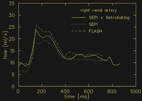
Figure 1 shows the results of the three different blood flow measurements in the right renal artery of a 31y old healthy volunteer. With all three techniques the typical flow pattern of an early systolic peak and a non-vanishing constant flow in diastole is observed. The different values for mean arterial flow are observed in this case are well within the range of physiologic variation. No systematic deviation was observed between the three measurement techniques.
With retrospective gating, a slightly improved signal-to-noise ratio was seen, as data are oversampled by a factor of 45/30 = 1.5. In total, image quality of retrospectively gated SEPI acquisitions was superior to SEPI with prospective gating, because data are acquired in a steady-state of the magnetization. In the future, retrospectively gated SEPI measurements in combination with contrast-enhanced MR angiographies might be able to provide a functional and morphologic evaluation of the renal arteries in only 3 breathholds.
1 SIEMENS Medical Systems, Erlangen, Germany
References
[1] Schönberg, S.O., Knopp, M.V., Bock, M., Kallinowski, F., et.al., Radiology 203, 45-53 (1997)
[2] Bock, M., Schönberg, S.O., Schad, L.R., et.al., JMRI 8, 889-895 (1998)
[3] Felblinger, J., Lehmann, C., Boesch, C., Magn. Reson. Med. 32, 523-529 (1994)