Sie befinden sich hier
Inhalt
Improved Target Volume Definition in Radiosurgery of Arteriovenous Malformations by Stereotactic Correlation of MRA, MRI, Blood Bolus Tagging, and Functional MRI
Lothar R. Schad, Michael Bock, Klaus Baudendistel, Marco Essig, Jürgen Debus, Michael V. Knopp, Rita Engenhart and Walter J. Lorenz.
Introduction
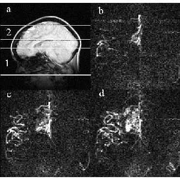
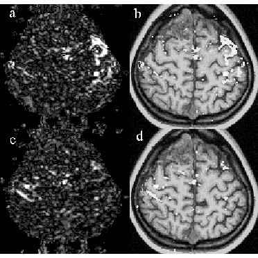
In a previous paper (1) we showed, that a helpful supplement to conventional planning of radiosurgery in patients with AVM is the correlation with MRA. The aim of the present study is a further improvement in the definition of the target volume (i.e. size of the nidus, the origin of feeding arteries, and the pattern of venous drainage) due to additional information about the haemodynamics of the AVM provided by dynamic MRA using a blood bolus tagging technique, whereas fMRI leads to an improved definition of structures at risk in patients with lesions close to the primary motor cortex area.
Results and conclusions
In combination to conventional MRI, MRA and 3D velocity phase-contrast imaging, information about the haemodynamics of the AVM was provided by a dynamic MRA with the STAR technique leading to an improved definition of the size of the nidus, the origin of the feeding arteries, and the pattern of the venous drainage (Fig. 1). In addition, fMRI was performed in patients with lesions close to the primary motor cortex area leading to an improved definition of structures at risk for the high dose application in radiosurgery (Fig. 2). Because of the inherent risk and discomfort of repeated conventional angiographic examinations, the different MR techniques are a useful completion in our treatment protocol for planning of radiosurgery and follow-up of AVM for studying the dynamic process of obliteration after radiation.
References
[1] Schad LR, Ehricke HH, Wowra B, Layer G, Engenhart R, Kauczor HU, Zabel HJ, Brix G, Lorenz WJ. Correction of spatial distortion in magnetic resonance angiography for radiosurgical planning of cerebral arteriovenous malformations. Mag Res Imag 1992;10(4):609-621.