Sie befinden sich hier
Inhalt
Correlation of Dynamic MRI- and PET-Data in Patients with Brain Tumors Receiving Radiotherapy
L. R. Schad, L. G. Strauss, A. Dimitrakopoulou-Strauss, M. V. Knopp, R. Engenhart
Introduction
Morphology and function can be evaluated and qualitatively as well as quantitatively assessed using MRI and PET in patients with brain tumors (1). In the last years dynamic MRI techniques have been developed to study tissue perfusion and vascular permeability by analyzing the Gd-DTPA kinetics within the framework of pharmacokinetic modeling (2). The aim of the present study is a direct correlation and proof of dynamic MRI-data with PET as a reference method for the assessment of the tissue perfusion with O-15 labeled water and the evaluation of regional F-18-Deoxyglucose (FDG) metabolism. This can be achieved by accurate image correlation using the stereotactic head frame.
Material and methods
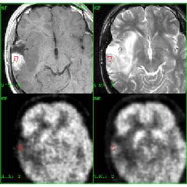
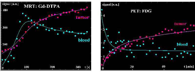
Five patients with astrocytomas were studied during the follow up after radiation therapy. MRI was performed to assess morphological (T1-, T2-weighted imaging) and functional (dynamic Gd-DTPA imaging) changes following radiotherapy. A fast FLASH imaging was used to receive functional information: TR = 170 ms, TE = 5 ms, FL = 90?, RFOV = 160x270 mm2, MA = 128x256, TH = 4 mm, 15 slices, acquisition time 22 s. The PET examination was performed using O-15 labeled water (1550 - 3600 Mbq, 4x30 s, 6x60 s) for the assessment of tissue perfusion. The regional metabolism was evaluated with FDG (240 - 360 Mbq, 20x60 s, 20x120 s) following the perfusion study. A dedicated mask in combination with a stereotactic localization system was used in MRI and PET for accurate and reproducible positioning of the patient. ROI’s were placed in identical MRI and PET brain areas for quantitative evaluation (Fig. 1). The signal-time curves have been parameterized with the help of a two compartment model (3) and the quantitative data of MRI and PET were compared (Fig. 2).
Results and conclusions
The comparison of MRI and PET demonstrate that Gd-DTPA is not correlated with the tissue perfusion as measured by PET. This is in accordance to the pharmacological properties of the substances. In contrast, a significant correlation was noted for the accumulation of Gd-DTPA and the regional FDG metabolism in tumors. When the Gd-DTPA accumulation and FDG metabolism in tumors is combined with the data for the normal brain tissue, no significant correlation was obtained. This result supports the theory, that the correlation between Gd-DTPA kinetics and the FDG metabolism is primarily based on the disruption of the blood-brain barrier, which is determining for the Gd-DTPA accumulation in MRI. With respect to this limitations we can conclude that MRI with Gd-DTPA reflects indirectly the active tumor regions in astrozytoma following radiation therapy.
References
[1] Schad LR, Boesecke R, Schlegel W, Hartmann G, Sturm V, Strauss L, Lorenz WJ. Three dimensional image correlation of CT, MR, and PET studies in radiotherapy treatment planning of brain tumours. J Comput Assist Tomogr 1987;11(6):948-954.
[2] Brix G, Semmler W, Port R, Schad LR, Layer G, Lorenz WJ. Pharmacokinetic parameters in CNS Gd-DTPA-enhanced MR imaging. J Comput Assist Tomogr 1991;15(4):621-628.
[3] Yokoi T, Iida H, Itoh H, Kanno I. A new graphic plot analysis for cerebral blood flow and partition coefficient with iodine-123-iodoamphetamine and dynamic SPECT validation studies using oxygen-15-water and PET. J Nucl Med 1993;34:498-505.