Sie befinden sich hier
Inhalt
Hyperpolarized 3He Magnetic Resonance Imaging of the Human Lung.
L. R. Schad1, P.Bachert1, M. Bock1, M. Essig1, M. Knopp1, M. Ebert2, T. Großmann2, W. Heil2, R. Surkau2, E. Otten2
Introduction
The use of hyperpolarized noble gases for magnetic resonance imaging (MRI) is of particular interest because of notorious limitations of standard proton MRI of the respiratory system. Accordingly, diagnostic imaging of the lungs is mainly performed by means of X-ray computed tomography (CT). Recently, MRI with hyperpolarized noble gas isotopes 129Xe and 3He has been applied to excised lungs of mouse (1) and to lungs of guinea pig (2), respectively. The purpose of our study is to establish 3He MRI in vivo in humans on a conventional whole-body MR scanner.
Material and method
Hyperpolarized 3He [spin 1/2, ? = 32.4 MHz/T, natural abundance 1.3x10⁻⁴] of high density was produced at the University of Mainz. The technique which is based on direct optical pumping of metastable 3He* atoms at low pressure by means of near-infrared laser light followed by electron-nuclear spin transfer allows to polarize ? 1018 helium atoms per second. The resulting spin polarization P(3He) ? 0.47 is 105 times larger than the thermal-equilibrium polarization of protons detected in standard MRI. Appropriate material for cell walls results in wall spin relaxation times T1 ? 100 h. Long T1 at room temperature allows easy storage and handling of polarized samples; e.g. a homogeneous magnetic field of a few Gauß is sufficient to maintain the enhanced 3He spin magnetization.
Hyperpolarized 3He MR imaging was performed at the German Cancer Research Center in Heidelberg on a whole-body MR scanner (Magnetom 63/84 SP 4000; Siemens, Erlangen, Germany) which is equipped with a rampable superconducting magnet of available field strengths of 0.1-2.0 T (Helicon, Siemens). We used different antenna systems originally developed for in vivo 31P MR spectroscopy at the standard field strength of 1.5 T in experiments at 0.8 T corresponding to 26.0 MHz Larmor frequency of 3He spins: a planar surface coil and a Helmholtz head coil with a loop diameter of 17 cm.
Results and conclusions
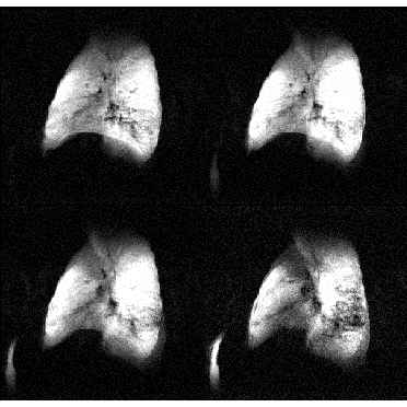
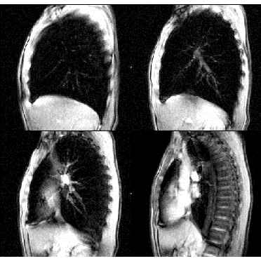
In vivo 3He FLASH images were obtained at 0.8 T from the lung of a volunteer following inhalation of hyperpolarized 3He gas. The volunteer was in supine position, its lung located within the sensitive volume of the Helmholtz coil. Following inhalation of and flushing with 4He gas, 3He was breathed in from a glass cell and a series of three times seven sagittal slices was obtained with a FLASH imaging technique in breathholding (Fig. 1). 3He images depict hollow spaces and anatomical structures of the lungs (informed consent was obtained from the volunteer prior to the MR examinations). After ramping the whole-body magnet back to the nominal field strength of 1.5 T conventional 1H FLASH imaging of the same anatomical region was performed (Fig. 2).
In conclusion, in vivo hyperpolarized 3He MRI is possible on a standard whole-body scanner providing a new modality lacking ionizing radiation for diagnostic imaging of the human respiratory system. MRI perfusion studies of the human lung will be possible with this high-sensitive non-proton MRI technique in the future.
1 Forschungsschwerpunkt Radiologie, Deutsches Krebsforschungszentrum, POB 101949, D-69009 Heidelberg
2 Institut für Physik, Johannes Gutenberg-Universität, D-55099 Mainz, Germany.
References
[1] Albert, M.S. et al. Biological magnetic resonance imaging using laser-polarized 129Xe. Nature 370, 199-200 (1994).
[2] Middleton, H. et al. MR imaging with hyperpolarized 3He gas. Magn. Reson. Med. 33, 271-275 (1995).