Sie befinden sich hier
Inhalt
M. Bock
Introduction
MR imaging of human lungs and airways is possible with hyperpolarized gases such as 3He [1]. Apart from the longitudinal relaxation time T1 two additional parameters determine the contrast of 3He images: the diffusion coefficient D and T2*. As D of 3He in the gas phase is 5 to 6 orders of magnitude larger than that of water, already the relatively weak imaging gradients of several mT/m create a significant diffusion weighting. In the human lung T2* is very short, because air-tissue boundaries induce rapid susceptibility changes.
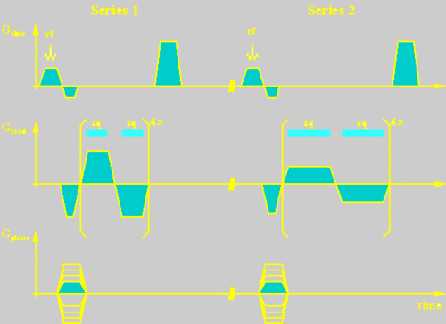
To measure these two parameters simultaneously in an acquisition time, that does not exceed the life-time of hyperpolarization in the human lung of about 30 s, a sequence was developed, that employs two gradient-echo trains with different inter-echo spacings and readout gradient strengths.
Materials and methods
The sequence for the simultaneous measurement of D and T2* was implemented on a clinical MR scanner (SIEMENS Magnetom 63/84 SP, Erlangen, Germany) using a home-built, double resonant 3He/1H Helmholtz coil (H.J. Zabel, DKFZ Heidelberg). Two gradient-echo trains with 8 alternating readout gradients each were sampled after a slice-selective rf excitation (Fig. 1). Readout gradient strength and bandwidth of the second echo train were half of that of the first to maintain spatial resolution (echo spacings: DTE1 = 4.56 ms, DTE2 = 7.12 ms, bandwidths: BW1 = 391 Hz/pixel, BW2 = 195 Hz/pixel).
The decay of the MR signal S during an echo train (normalized to the signal of the first echo S(0)) is described by

with
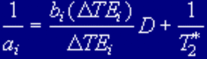
where n = 0..7 is the echo number and i = 1 or 2 is the series index. As the b-value bi(DTEi) is dependent on the details of the gradient switching, it is different for series 1 and 2. Values of D and T2* are calculated from the decay constants ai, which are determined by a fitting procedure.
Measurements with both hyperpolarized and non-prepolarized gas samples of 3He in glass cells were performed. Hyperpolarization of about 60% was created at the University of Mainz (Group: E. W. Otten) by laseroptical pumping of metastable 3He with subsequent collisional polarization transfer to the ground state [2]. MR imaging was performed using very low flip angles (a < 1°) to preserve hyperpolarization (Matrix / FOV / SL / NEX = 96-128 / 500 mm / 40 mm / 1). The non-prepolarized sample contained a 3He/O2 gas mixture in a sealed cell (T1 = 2.5 s). At TR = 100 ms flip angles around the Ernst angle aE = 16° were chosen. The other imaging parameters were: Matrix / FOV / SL / NEX = 642 / 300 mm / 40 mm / 1. For comparison, 3He diffusion coefficients were also measured with a spectroscopy sequence.
Results and discussion
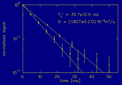
Fig. 2 shows the result of a measurement with hyperpolarized 3He. The measured value of D is in good agreement with the spectroscopy measurement (1.87× 10⁻⁴ m2/s) and the literature value (1.83×10⁻⁴ m2/s, T = 20°C, p = 1 bar). In general it was observed, that T2*-values showed larger uncertainties than values of D.
From the 2 image series maps of D and T2* can be calculated. Changes in the structure of lung tissue modify local diffusion coefficients and T2*. Parameter maps derived from 3He imaging data might help to identify regions in the human lung, that are affected by pathologic processes.
References
[1] Bachert P., Schad L.R., Bock M., et.al.: Magn. Reson. Med. 36, 192-196 (1996)
[2] Eckert G., Heil W., Meyerhoff M., et. al.: Nucl. Instrum. Meth. A 346, 45-51 (1994)