Sie befinden sich hier
Inhalt
T2-Measurments for Polymer Gel Based Three-dimensional Dosimetry
J. Ahlswede, M. Bock, T. Bortfeld, K.-H. Hoever, F. Oberdorfer, L. R. Schad
Introduction
In modern radiology complicated three-dimensional dose distributions are used for tumour treatment. This is possible due to the development and improvement of radiosurgery systems including multi-leaf-collimators and brachytherapy. For clinical implementation it is necessary to evaluate exact dose distributions. Ionisation chambers can only measure integral dose of a volume and film dosimeters are able to measure dose in a plain, both being standard dosimeters. Therefore, it is time consuming to verify three-dimensional dose distributions.
Polymer gel dosimeters on the other hand do provide dose distributions in three dimensions. Polymer gels are easy to handle, formable and stabile while handling.
Methods and materials
In polymer gel dosimetry advantage is taken of the physical and chemical properties of monomers embedded in a gel matrix. Radiation induces polymerisation of the monomers forming polymer chains. This reduces the spin-spin-relaxation time T2 of the hydrogen atoms in the irradiated area and makes the effect accessible to MRI. Within a certain dose range the change in the relaxation rate R2 = 1/T2 is directly proportional to the applied dose. Usually, T2-maps are taken by a Carr-Purcell-Meiboom-Gill sequence (CPMG) where a series of T2-weighted images is obtained at different echo times TE.
In this study home-made polymer gels and gels from MGS Inc. (Guilford, USA) have been used. For sensitivity measurements the gels have been irradiated by linear accelerators (Mevatron, Siemens, Germany) with photon energies of 6 and 15 MeV and also a 60Co source with 1.3 MeV energy was used (Gammatron, Siemens, Germany). During treatment different collimators and doses have been applied for obtaining spatial as well as dose resolution. To test other radiation sources the gel was irradiated by a neutron beam with a maximum energy of 32 MeV.
Finally polymer gel dosimeter were compared with X-OMAT V film dosimeters (Kodak, Germany), where the dose distributed to the gel was five times larger than the one the film was irradiated with.
After irradiation the gel was measured at a clinical 1.5T whole body scanner (Magnetom SP, Siemens, Germany). A 32-echo Carr-Purcell-Meiboom-Gill sequence (CPMG) was used to acquire T2-maps. Typical parameters are: TE = 15 - 1000 ms; TR = 2 - 5 s; FOV = 220 - 250 mm; matrix size = 128x128 to 512x512. T2-maps were calculated pointwise by a linear fit of the logarithmic signal amplitudes. The temporal stability was verified by taking T2-maps throughout a period of six months after irradiation.
Results and discussion
Sensitivity measurements showed a linear dependency between dose and R2, the independence of beam energy and radiation source as published by MGS [1 – 3]. The comparison with film dosimetry showed a comparable resolution in both techniques. The stability measurements showed a strong shortening in T2 values over time, which can be explained by oxygen penetration into the gel after irradiation causing additional polymerisation. Nonetheless, the irradiated areas could still be separated from unirradiated areas.
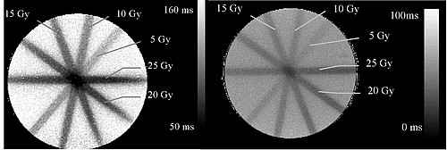
right: T2-map of the same gel 22 weeks after irradiation.
In the future, the CPMG sequence will be improved to increase spatial resolution, T2 calculation and measure three-dimensional dose distributions. Further developments of the pulse sequence will focus on optimisation of handling, dose verification and time saving Therefore the clinical utility for quality assurance in radio therapy will be evaluated in selected treatment plans.
References
[1] Maryanski, M.J. et al, [1994] Phys. Med. Biol. 39: 1437-1455.
[2] Maryanski, M.J. et al, [1996] Med. Phys. 23(5): 699-705.
[3] Ibbott,G .et al, [1997] Int.J. Radiat. Oncol.Biol.Phys. 38:1097-1103.