Sie befinden sich hier
Inhalt
Comparison of TurboFLASH and Spin-Echo-R1-Measurements of FeMRI-Gel-Phantoms for verification of 3D-dose distributions
A. W. Hartlep, M. Bock, F. Oberdorfer, L. R. Schad
Introduction
Since stereotactic radiation therapy has become increasingly important in brain tumour therapy, there is a need to verify the delivered complex dose distributions. FeMRI dosimetry is able to confirm any spatial dose distribution. Like the conventional Fricke dosimetry FeMRI is based on the radiation induced oxidation of Fe2+ to Fe3+. The amount of ferric ions depends linearly on the absorbed radiation dose [1]. If an aqueous Fricke solution is infused in a gelatine matrix, a three-dimensional dose distribution can be detected by measuring the spatial concentration of ferric ions. Gore et al. introduced a NMR technique based on the effect of different spin-lattice relaxation rates (R1) of protons in presence of ferrous or ferric ions.
To improve the ability for clinical use of the FeMRI-Gel-dosimetry the reproducibility has to be studied as well as the precision of this method. The crucial point is the R1-measurement of the irradiated gel. Two different R1 measurement techniques were compared in this study.
Materials and methods
The Fricke-Gel used consisted of 7.5% gelatine by weight and a ionic solution that was composed of NaCl (1 mM), (NH4)2Fe(SO4)2 (1 mM) and H2SO4 (0.05 M) relative to the total volume. The pH level was found to be about one and the density of the FeMRI gel was 1.005 g cm-3. The gel was filled in twelve calibration vials of 20 cm3 and in a glass head phantom of 16 cm diameter. Calibration vials were irradiated with discrete doses from 0 to 30 Gy while the head phantom was irradiated with four coplanar conformal beams using a MEVATRON KD2™ (Siemens) linear accelerator at a photon energy of 15 MV.
Immediately after irradiation the T1 values were measured at a MAGNETOM 63/64 SP™ (Siemens) MR-scanner at 1.5 T using a standard head coil. Two different sequences were used to acquire T1-images: a conventional Spin-Echo-sequence (SE) and a TurboFLASH (TFL) technique [2]. For the SE-method two pictures with repetition times of TR1 =
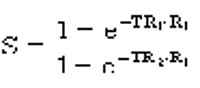
2900 and TR2 = 700 ms were taken. The R1-value of each pixel was calculated from the quotient S of the gray values of both pictures:
For the TFL technique 25 pictures with different inversion times (TI) between 100 and 4000 ms were taken and the R1-value was calculated from a 3-parameter fit using the equation:

The parameters A0, NRF = (1-cosß)/2 and T1 were fitted. The whole SE and TFL measurements were repeated five times and the accuracy was compared. With the obtained parameters the gels were calibrated and the T1-values of the head phantom can be converted to doses and a dose picture was calculated.
Results and Discussion
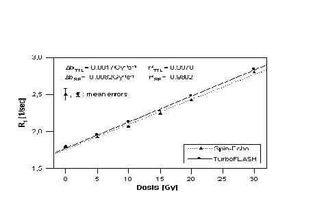
Fig. 1 shows the results of the calibration measurements using SE and TFL images. The plot of the TFL measurement gives a linear relationship with a slope b of bTFL = (0.0343 ± 0.0017) s-1Gy-1 and a linear dependence of the values of r²TFL = 0.9970. The slope of the SE-measurement is bSE = (0.0334 ± 0.0082) s-1Gy-1 and the regression coefficient is r²SE = 0.9803.
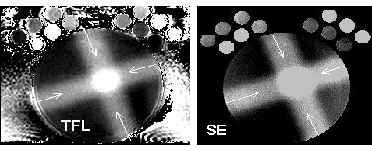
Since the calculated dose in a gel phantom is proportional to b-1 the dose is very sensitive to slope errors. The slope error ?b has to be minimized to improve the accuracy of the FeMRI-dosimetry. Taking into consideration that ?bTFL is about five times smaller than ?bSE these results indicate a higher reproducibility of the TFL method over the SE technique. In order to get reliable quantitative results, the TFL technique should be preferred to the SE method. As Fig. 2 shows TFL pictures are prone to artefacts because of the non-ideal point-spread-function. SE images give a better visualisation of spatial dose distributions. So the SE technique should be preferred for qualitative demonstrations and the TFL method for quantitative predictions.
References
[1] Gore J.C., Kang Y.S., Schulz R.J.: Phys. Med. Biol. 29, 1189-1197 (1984)
[2] Blüml S., Schad L.R., Stepanow B., et. al.: Magn. Reson. Med. 30, 289-295 (1993)