Sie befinden sich hier
Inhalt

Neuroanatomy
Professor of Neuroanatomy
Team Members:
Dr. Nina Bonekamp
Dr. Maren Engelhardt
Dr. Femke Groeneweg
PD Dr. Markus Islinger
Plasticity of neuronal compartments and organelles
Neurons are highly polarized cells that have clearly distinguishable subcellular compartments. These compartments have characteristic structural and functional properties. The main neuronal compartments consist of the somatodendritic, axonal and synaptic domains. Our studies mainly focus on the structural plasticity of the axon. We primarily investigate the molecular composition and structural plasticity of the so-called axon initial segment (AIS).
Our main research interests are
- Axonal plasticity and development of the axon initial segment in vivo
- Neuronal polarity
- Contact between membranes of organelles
- Role of peroxisomes in neurodegeneration
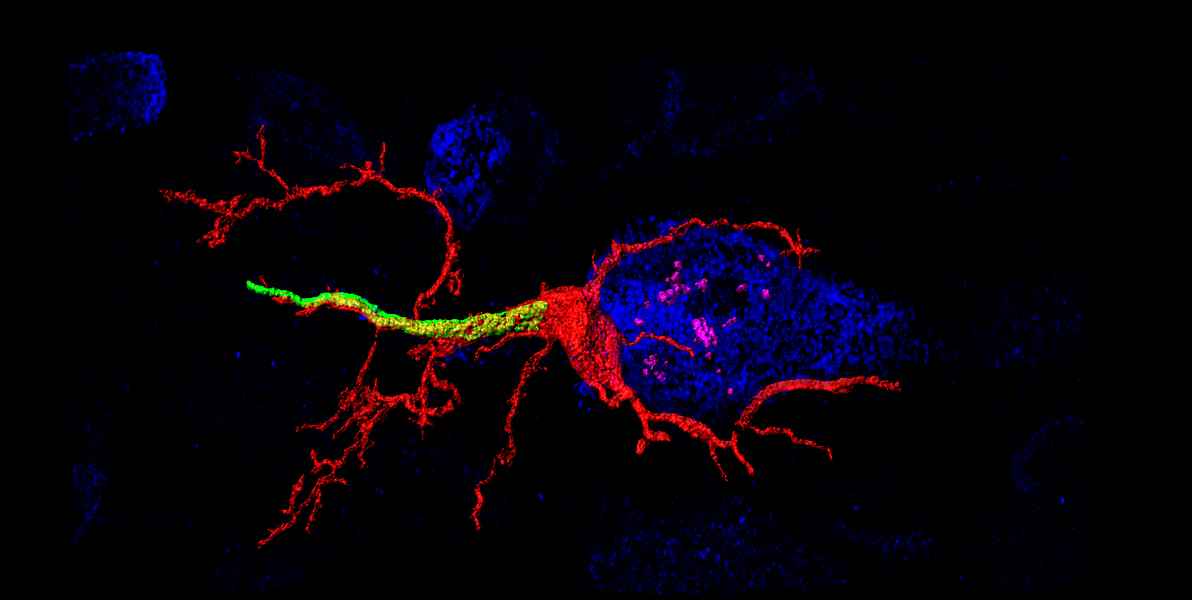
The AIS corresponds to the first segment of the axon and is of crucial functional importance as the primary trigger zone for the action potential. Current research data show an amazingly strong plasticity of the AIS: Both the molecular composition and the length and position of the AIS are dynamically regulated and contribute to the regulation of neuronal excitability. In this context, we want to further elucidate the molecular basis of structural AIS plasticity - especially in the developing nervous system. Furthermore, we are investigating the mechanisms underlying the directed transport of cell organelles. This second research approach of our group aims to understand the role of membrane contacts between peroxisomes and the endoplasmic reticulum (ER) in neurons.
Recent studies show that both structure and function of AIS are dynamically regulated. One of our working hypotheses assumes that this plasticity contributes to the intrinsic excitability of neurons. Using the mouse whisker-to-barrel pathway in vivo, we have shown recently that AIS plasticity is a homeostatic, bidirectional process, which is reflected by changes in AIS length and neuronal excitability. We further uncovered two distinct timescales, in which AIS plasticity occurs in vivo. For example, the remodeling of AIS length, accompanied by significant changes in neuronal excitability, was observed within 1 hour of exposure of the animal to an enriched environment. We now aim at understanding the role of AIS plasticity within the physiological range of sensory processing and will utilize a new live reporter mouse line to begin in vivo AIS imaging studies in awake and behaving animals.
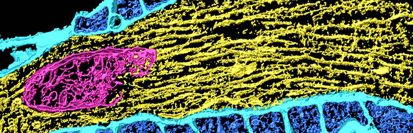
Contact sites between the membranes of peroxisomes and the endoplasmic reticulum have been shown on an ultrastructural level for quite some time. These contact sites are probably of great importance for a large number of physiological processes and the cell, including neuronal transport, intracellular lipid metabolism and the exchange of phospholipids between adjacent biomembranes. Recently, we succeeded in identifying the peroxisomal membrane protein (acyl-CoA binding protein 5; ACBD5) as a new binding partner, the ER-associated membrane protein VAPB. This was the first time that we were able to clarify the molecular basis for establishing membrane contacts between peroxisomes and ER.
In the future, the Department of Neuroanatomy will focus on the following topics:
- Development and plasticity of axon-carrying dendrites (AcD) in pyramidal neurons
- Recruitment of AcD cells into functioning hippocampal and cortical ensembles
- Untangling AIS plasticity in a reporter mouse model in vivo
- Functional relevance of membrane contacts between ER and PO
- Functional role of peroxisomes for neurodegenerative changes in the retina
Selected publications
- Cell lineage-specific mitochondrial resilience during mammalian organogenesis.
Burr SP, Klimm F, Glynos A, Prater M, Sendon P, Nash P, Powell CA, Simard ML, Bonekamp NA, Charl J, Diaz H, Bozhilova LV, Nie Y, Zhang H, Frison M, Falkenberg M, Jones N, Minczuk M, Stewart JB, Chinnery PF. Cell. 2023. 186(6):1212-1229.e21. doi: 10.1016/j.cell.2023.01.034 - Dendritic axon origin enables information gating by perisomatic inhibition in pyramidal neurons.
Hodapp A, Kaiser ME, Thome C, Ding L, Rozov A, Klumpp M, Stevens N, Stingl M, Sackmann T, Lehmann N, Draguhn A, Burgalossi A, Engelhardt M, Both M. Science. 2022. 377(6613):1448-1452. doi: 10.1126/science.abj1861 - PEX11β and FIS1 cooperate in peroxisome division independently of mitochondrial fission factor. Schrader TA, Carmichael RE, Islinger M, Costello JL, Hacker C, Bonekamp NA, Weishaupt JH, Andersen PM, Schrader M. J Cell Sci. 2022. 135(13):jcs259924. doi: 10.1242/jcs.259924
- Starting the engine of the powerhouse: mitochondrial transcription and beyond.
Miranda M, Bonekamp NA, Kühl I. Biol Chem. 2022. 403(8-9):779-805. doi: 10.1515/hsz-2021-0416. - Sensory input drives rapid homeostatic scaling of the axon initial segment in mouse barrel cortex.
Jamann N, Dannehl D, Lehmann N, Wagener R, Thielemann C, Schultz C, Staiger J, Kole MHP, Engelhardt M. Nat Commun. 2021. 12(1):23. doi: 10.1038/s41467-020-20232-x - Cerebellar and hepatic alterations in ACBD5-deficient mice are associated with unexpected, distinct alterations in cellular lipid homeostasis.
Darwisch W, von Spangenberg M, Lehmann J, Singin Ö, Deubert G, Kühl S, Roos J, Horstmann H, Körber C, Hoppe S, Zheng H, Kuner T, Pras-Raves ML, van Kampen AHC, Waterham HR, Schwarz KV, Okun JG, Schultz C, Vaz FM, Islinger M. Commun Biol. 2020. 3(1):713. doi: 10.1038/s42003-020-01442-x - The diversity of ACBD proteins - From lipid binding to protein modulators and organelle tethers.
Islinger M, Costello JL, Kors S, Soupene E, Levine TP, Kuypers FA, Schrader M. Biochim Biophys Acta Mol Cell Res. 2020. 1867(5):118675. doi: 10.1016/j.bbamcr.2020.118675 - Dynamic Regulation of Synaptopodin and the Axon Initial Segment in Retinal Ganglion Cells During Postnatal Development.
Schlüter A, Rossberger S, Dannehl D, Janssen JM, Vorwald S, Hanne J, Schultz C, Mauceri D, Engelhardt M. (2019) Front Cell Neurosci. 13:318. doi: 10.3389/fncel.2019.00318 - Intracellular redistribution of neuronal peroxisomes in response to ACBD5 expression.
Wang Y, Metz J, Costello JL, Passmore J, Schrader M, Schultz C, Islinger M. PLoS One. 2018.13(12):e0209507. doi: 10.1371/journal.pone.0209507 - Structural Plasticity of Synaptopodin in the Axon Initial Segment during Visual Cortex Development.
Schlüter A, Del Turco D, Deller T, Gutzmann A, Schultz C, Engelhardt M. Cereb Cortex. 2017. 27(9):4662-4675. doi: 10.1093/cercor/bhx208
Photo Credits: #1 FGV-Zentrum, Med. Fakultät Mannheim
Kontextspalte
Contact
Medical Faculty Mannheim
Heidelberg University
Ludolf-Krehl-Str. 13-17
68167 Mannheim
Deutschland
Phone +49 621 383-71555
christian.schultz@medma.uni-heidelberg.de