Sie befinden sich hier
Inhalt

Head of the Department of Neuroradiology
Professor for Neuroradiology
Experimental Neuroradiology and preclinical imaging
Team members:
PD Dr. Holger Wenz
Preclinical research is a key step in the development of new treatment methods and precedes clinical research on humans. The aim of the research group is therefore to use experimental models with the representation of both physiological and pathological processes to determine new methods and active substances to a precisely defined treatment goal.
The research focus of the group is CT-based small animal imaging and imaging of various aspects of neuro-oncology. We cooperate with other research groups, which also offers the possibility of MRI-based (1 Tesla small animal MRI; Bruker BioSpec 94/20) and molecular imaging.
A volume CT (Yxlon Fox) is available for experimental and small animal imaging. Due to a flexible positioning of the object to be examined in the beam path, the device allows a very high geometric magnification. Depending on the size of the sample, a very high resolution down to the single-digit micrometer range can thus be achieved. The conical beam geometry of the beam path allows a larger volume to be acquired with only one rotation of the object, which significantly reduces acquisition time. A fast examination protocol (so-called Quick Scan) can acquire CT images within an examination time of 33 seconds with up to 1000 projections. Proportionally shorter examination times are possible with a reduced number of projections.
An example of the importance of preclinical imaging is the research of subarachnoid hemorrhage (SAH) and its consequences. With SAH, a life-threatening and very painful bleeding into the subarachnoid space occurs spontaneously. The most frequent cause of this is the rupture of an aneurysm. In the further course of the disease, up to 70% of patients experience constriction of the blood vessels supplying the brain, so-called cerebral vasospasm, despite initial successful treatment of the source of bleeding. The constriction of the vessels leads to reduced perfusion and further cerebral damage, which is often accompanied by permanent damage and might be fatal.
With our volume CT we are able to perform high-resolution digital subtraction angiography (DSA) of the cerebral vessels in laboratory rodents (Fig. 1).
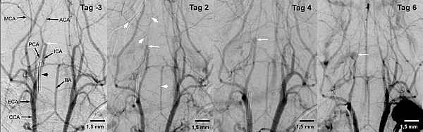
This examination allows the evaluation of changes in the diameter of cerebral vessels following experimentally induced subarachnoid hemorrhage in a longitudinal course. In the further course, there is the possibility to test different therapeutic approaches and to investigate their effects.
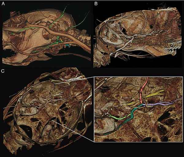
In addition, digital subtraction angiographies can be made in the thoracic and abdominal area. A further option for imaging intracerebral vessels is the use of Quick Scan to produce high-resolution CT images of the brain vessels of the mouse. (Fig. 2).
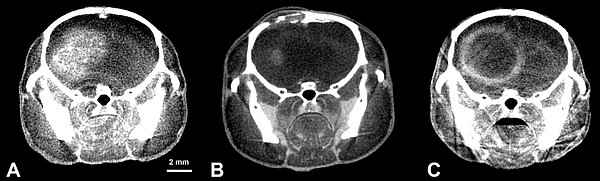
In addition to questions on the vascular status, oncological issues are also being addressed, such as the visualization of the smallest liver metastases in mice after application of a nanoparticulate contrast agent (ExiTtron nano 6000; nanoPET Pharma GmbH, Berlin, Germany). Furthermore, intracerebral tumors, such as glioblastoma multiforme, can be well visualized after application of a contrast agent (Iomeprol, Imeron 300) (Fig. 3).
Preclinical imaging can be used to investigate the longitudinal course of glioblastomas and other tumor types and the effect of potential new drugs in animal models. Moreover, it is possible to generate a needle beam by collimation of the cone beam in the micro-CT, with which a precise irradiation of the brain tumor is possible.
A further focus of the research group is the ex vivo imaging of human and animal preparations as well as various medical devices. These include stents that are tested for implantation in humans to exclude possible damage. As well as imaging of insects that have been stained with X-ray contrast agent. It is also possible to examine already extracted teeth, which, due to the different X-ray densities of their structures, allow a distinction to be made between enamel, dentine and pulp cavity, thus allowing micro-fissures or changes in the enamel to be detected. Furthermore, bones from experimental fracture models can be assessed with regard to fracture healing (callus formation, trabecular thickness).
Cooperation partners:
- Prof. Dr. med. Marc. A. Brockmann, Klinik und Poliklinik für Neuroradiologie, Universitätsmedizin Mainz
- Priv.-Doz. Dr. med. Frank A. Giordano, Klinik für Strahlentherapie und Radioonkologie, Universitätsmedizin Mannheim
- Prof. Dr. med. Dr. rer. nat. Lothar Schilling, Abteilung für Neurochirurgische Forschung, Universitätsmedizin Mannheim
- Prof. Dr. B. Wängler, Molekulare Bildgebung und Radiochemie, Institut für klinische Radiologie und Nuklearmedizin, Universitätsmedizin Mannheim
- Prof. Dr. med. Daniel Nowak, III. Medizinische Klinik für Hämatologie und Onkologie, Universitätsmedizin Mannheim
Selected publications
- Comparative Analyses of the Impact of Different Criteria for Sepsis Diagnosis on Outcome in Patients with Spontaneous Subarachnoid Hemorrhage.
Centner FS, Oster ME, Dally FJ, Sauter-Servaes J, Pelzer T, Schoettler JJ, Hahn B, Fairley AM, Abdulazim A, Hackenberg KAM, Groden C, Etminan N, Krebs J, Thiel M, Wenz H, Maros ME. J Clin Med. (2022) 11(13):3873. - Comparative analysis of machine learning algorithms for computer-assisted reporting based on fully automated cross-lingual RadLex mappings.
Maros ME, Cho CG, Junge AG, Kämpgen B, Saase V, Siegel F, Trinkmann F, Ganslandt T, Groden C, Wenz H. Sci Rep (2021) 11(1):5529. - Longitudinal imaging and evaluation of sah-associated cerebral large artery vasospasm in mice using micro-ct and angiography.
Weyer V, Maros ME, Kronfeld A, Kirschner S, Groden C, Sommer C, Tanyildizi Y, Kramer M, Brockman MA. J Cereb Blood Flow Metab (2020) 40(11):2265-2277. - Imaging of orthotopic glioblastoma xenografts in mice using a clinical ct scanner: Comparison with micro-ct and histology.
Kirschner S, Murle B, Felix M, Arns A, Groden C, Wenz F, Hug A, Glatting G, Kramer M, Brockmann MA. PloS One. (2016) 11(11):e0165994. - A low cost metal-free vascular access mini-port for artifact free imaging and repeated injections in mice.
Fiebig T, Figueiredo G, Boll H, Kerl HU, Noelte IS, Forster A, Groden C, Brockmann MA. PloS One (2013) 8(6):e65939. - In vivo x-ray digital subtraction and ct angiography of the murine cerebrovasculature using an intra-arterial route of contrast injection.
Figueiredo G, Boll H, Kramer M, Groden C, Brockmann MA. AJNR Am J Neuradiol (2012) 33(9):1702-9. - Comparison of digital subtraction angiography, micro-computed tomography angiography and magnetic resonance angiography in the assessment of the cerebrovascular system in live mice.
Figueiredo G, Brockmann C, Boll H, Heilmann M, Schambach SJ, Fiebig T, Kramer M, Groden C, Brockmann MA. Clin Neuradiol (2012) 22(1):21-8. - Three-dimensional in vivo imaging of the murine liver: A micro-computed tomography-based anatomical study.
Fiebig T, Boll H, Figueiredo G, Kerl HU, Nittka S, Groden C, Kramer M, Brockmann MA. PloS One (2012) 7(2):e31179. - Vascular imaging in small rodents using micro-ct.
Schambach SJ, Bag S, Groden C, Schilling L, Brockmann MA. Methods (2010) Jan;50(1):26-35. - Ultrafast high-resolution in vivo volume-cta of mice cerebral vessels.
Schambach SJ, Bag S, Steil V, Isaza C, Schilling L, Groden C, Brockmann MA. Stroke (2009) 40(4):1444-50.
Photo Credits: #1 FGV-Zentrum - Med. Fakultät Mannheim
Kontextspalte
Contact
Medical Faculty Mannheim
Heidelberg University
Theodor-Kutzer-Ufer 1-3
68167 Mannheim
Phone +49 621 383-2443
neuroradiologie@umm.de