Sie befinden sich hier
Inhalt
M. Amann, M. Bock, L. R. Schad
Introduction
The time-of-flight magnetic resonance angiography (TOF-MRA) has been validated as a non-invasive screening technique in many applications, for example of the intracranial vasculature [1] and the renal arteries [2]. Often TOF-MRA is combined with 3D excitation techniques to gain a higher signal-to-noise (S/N) ratio and isotropic resolution. The advantage that TOF-MRA does not use any contrast agent is obscured by the fact that conventional TOF-MRA suffers from long acquisition times (usually in the range of 5-10 minutes). This is crucial in the case of renal angiography where the attainable resolution is limited by respiratory motion.
Spiral MRI offers some advantages to angiography in comparision to techniques with cartesian readout. Due to the attainable short echo times and the intrinsic compensation of higher gradient moments, spiral MRI is less sensitive to flow artifacts [3]. Additionally, spiral MRI samples the k-space in a very efficient way, so it is a promising technique to shorten acquisition time, thus allowing measurements in a single breathhold.
Material and methods
To investigate the benefit of spiral readout, spiral TOF-MRA of the intracranial vessels and of the renal arteries were performed. The sequences were implemented on a 1.5 T whole body scanner (Magnetom Vision, Siemens, Germany). In both cases a conventional phase-encoding gradient in partition direction was combined with a inplane spiral readout. The spiral trajectories were slewrate-optimized with respect to the chosen number of interleaves and to the selected field-of-view (FOV) [4]. The 3D slab was excited with a TONE (tilted optimized nonsaturating excitation) pulse [5], resulting in a linear varying flip angle over the slab. The mean angle was chosen to be 20° in both cases. In the case of less time-critical brain measurements, the excitation pulse was preceded by a magnetisation transfer (MT) pulse [6] to saturate the stationary tissue. A frequency selective pulse was additionally applied to presaturate fat protons. 32 interleaved spirals with a duration of 15.36 ms and 2048 k-space samples were acquired per partition. The other parameters were: 44 partitions with a thickness of 1.5 mm, TR/TE = 41.5 ms/2.0 ms. The resulting acquisition time was one minute.
To reduce acquisition time for the renal angiography no presaturation pulses were applied. The duration of a single spiral was reduced to 7.68 ms to prevent severe artifacts caused by off-resonant spins. To achieve comparible in-plane resolution, bandwidth and S/N ratio, 64 interleaved spirals with 1024 data points were used. The other parameters were: TR/TE = 10.4 ms/1.3 ms. An asymmetric k-space sampling and sinc-interpolation in partition direction halves the acquisition time providing comparable resolution. For a 60 mm slab with 40 interpolated partitions an acquisition time of 13 s was obtained.
For both MRA measurements maximum intensity projections (MIP) were calculated. The MIP images of the renal arteries were computed on a volume as small as possible to gain optimal vessel contrast ('targeted MIP').
Results and conclusions
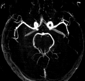
Fig. 1 shows the axial MIP of a spiral TOF intracranial MRA. At a FOV of 250x250 mm2 an inplane resolution of 0.68 mm was obtained. Though blurring of fine structures caused by B0 inhomogeneities reduces the theoretical resolution, even small vessels are clearly visible.
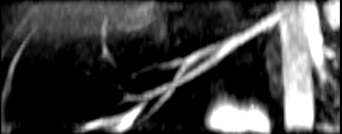
Fig. 2 shows the targeted MIP of a spiral TOF-MRA representing the right renal artery. An inplane resolution of 0.97 mm was achieved at a FOV of 400x400 mm2. Even intrarenal branches (arrows) are shown.
In this work we demonstrated the efficiency and quality of spiral TOF-MRA. Offline correction with respect to the effects of B0 inhomogeneities will further improve image quality. With this sequence scheme it is now possible to get renal TOF angiograms within a single breathhold. The quality of the angiograms acquired from healthy volunteers will now be verified in screening pathological processes like arterial stenosis or arteriovenous malformations. The combination of spiral MRA with contrast agent to enhance vessel contrast will be an additional focus of future work.
References
[1] Marchal G. et al., Radiology 171, 793-799, 199
[2] Fellner C. et al., Radiology 196, 681-687, 1995
[3] Van Tyen R., Saloner D., 6th ISMRM, 779, 1998
[4] King K., Foo T., Crawford C., MRM 34, 156-160, 1995
[5] Atkinson D. et al., Radiology 190(3), 890-894, 1994
[6] Pike G. et al., MRM 25, 372-379, 1992