Sie befinden sich hier
Inhalt
Computer Assisted Clinical Medicine
Professor of Medical Physics
Medical Physics, Imaging
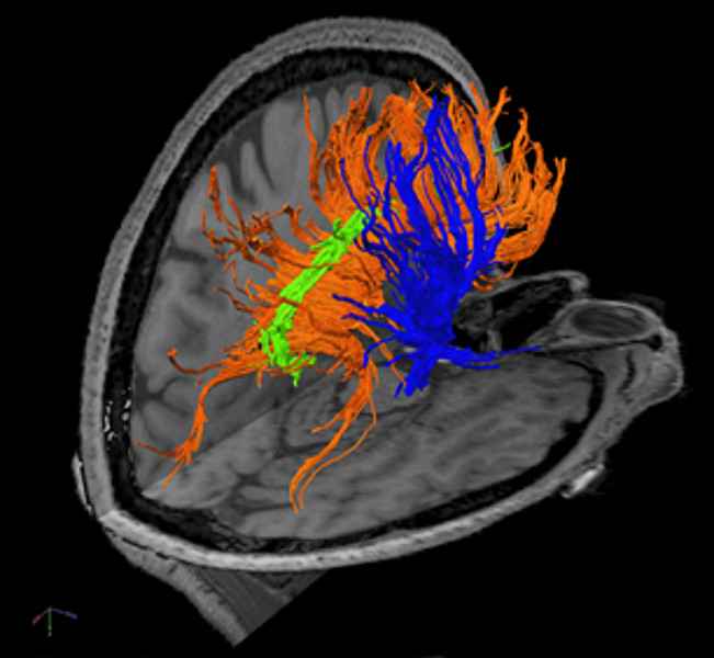
We are working on several aspects of improved oncological radiotherapy treatment planning and monitoring by using physiological and functional imaging of CT, MRI and PET. One of our main research aspects lies in developing new MR techniques (23Na imaging, dynamic MRT, diffusion, perfusion, blood bolus tagging, BOLD MRI) for clinical use in therapy planning and monitoring. Another main aspect denotes imaging of hyperpolarized 3He in the human lung, as well as T2*- and T1-techniques for non-invasive measurement of tissue oxygenation and perfusion in the myocardium which is of general interest in radiology.

Major Contributions
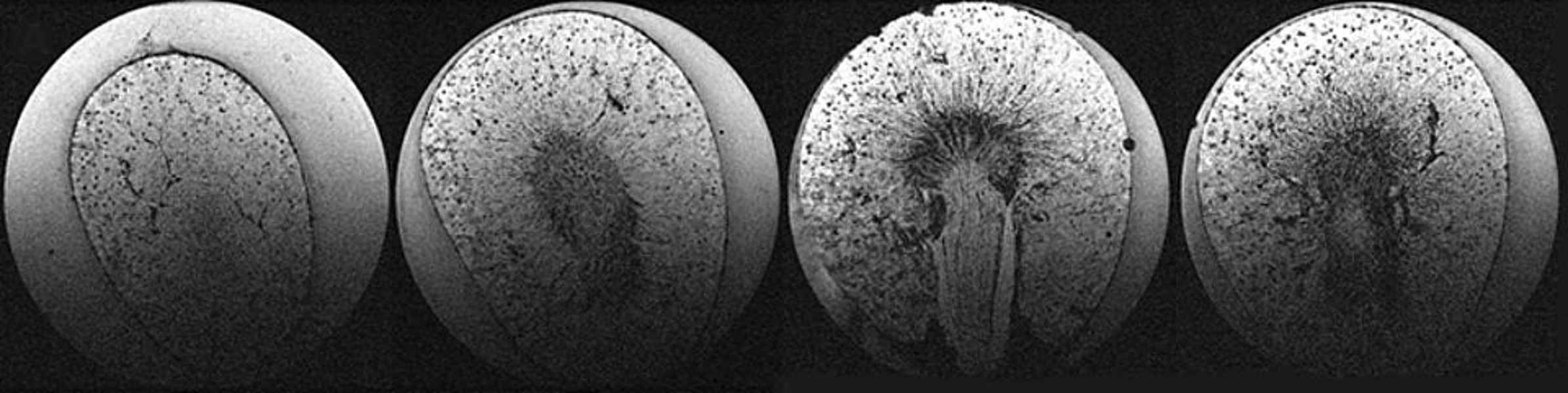
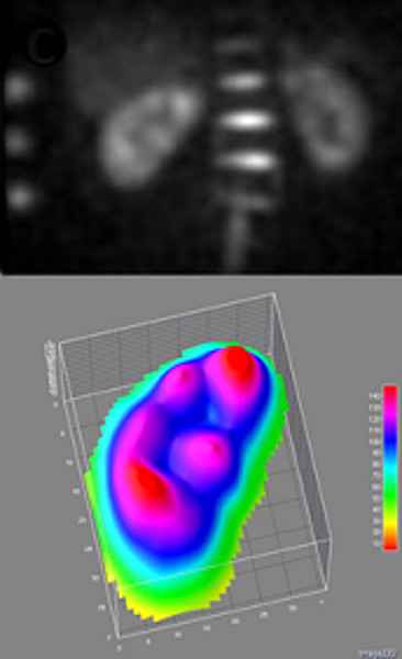
Our overall goal is the evaluation of quantitative biomarkers for diagnosis and/or therapy monitoring accomplished by translational animal/cell experiments. Major contributions are the evaluation of glomeruli counts in rat kidneys using MR at 9.4 T; the establishment of a high-resolution protocol for diffusion tensor imaging at 3 T for tracking fibers in the brain and the clinical evaluation of diffusion kurtosis in whole body applications; the development of an MR set up for measuring sodium concentration in human kidneys at 3 T; the implementation of a protocol for use in clinical contexts for mapping of perfusion and ventilation in the human lung at 1.5 T; and the development of an MR set up to measure perfusion maps in the human kidney for quantitative evaluation at 3 T.

What we want to know
We do have many years of experi¬ence (since 1985) in developing new methods and techniques for magnetic reso¬nance imaging with strong focus on Medical Technology using MR imaging and spectroscopy for modern treatment planning and monitoring. The institute is equipped with 3x 1.5T and 2x 3T whole-body MR systems and has also access to a 9.4T animal system at the Central Institute of Mental Health.
Besides, we are developing molecular innovative imaging technologies by fusion of several imaging modalities (CT, MRI, PET) to enable image-guided, high-precision interventions using high-end CT and robotic systems (ZEEGO, Siemens). The group is composed of scien¬tists from physics, electrical engineering, and computer science and is working in close co-operation with the local medical departments (Radiology, Radiotherapy, Neuroradiology, Neurology and Cardiology). The aim of the research is to design innovative diagnostic methods and procedures to optimize morphologi¬cal, functional and metabolic diagnosis as a basis for radiology and image-guided radiotherapy, as well as to establish an experimental clinical translational research using high-resolution animal imaging.
The focus of the current development work are methods for physiological MR imaging (perfusion, diffusion, oxygenation) on hu¬mans especially in the abdomen (kidney, lung, heart), as well as sodium imaging for non-invasive measurement of tissue viability. A further objective is the establishment of animal models (rat and mouse) for building various translational studies (high-resolution morphology, X-nuclear imaging, stroke model and renal glomeruli representa¬tion) accomplished by cell experiments in a bioreactor system.
National and International Joint Research Projects
BMBF M2OLIE
DFG-Radiomics ZO 257/8-1
DFG-FDLunge ZO 257/6-1
DFG-Radiomics RectumCA ZO 257/9-1
Selected Publications
- 23 Na MRI in ischemic stroke: Acquisition time reduction using postprocessing with convolutional neural networks.
Adlung A, Paschke NK, Golla AK, Bauer D, Mohamed SA, Samartzi M, Fatar M, Neumaier-Probst E, Zöllner FG, Schad LR. NMR Biomed (2021) Apr;34(4):e4474. - Prediction of peripheral nerve stimulation thresholds of MRI gradient coils using coupled electromagnetic and neurodynamic simulations.
Davids M, Guérin B, vom Endt A, Schad LR, Wald LL. Magn Reson Med (2019) 81(1):686-701. - Feasibility study of a double resonant 8-channel 1H/ 8-channel 23Na receive-only head coil at 3 Tesla.
Malzacher M, Chacon-Caldera J, Paschke N, Schad LR. Magn Reson Imaging (2019) 59:97-104. - Na Triple-quantum signal of in vitro human liver cells, liposomes, and nanoparticles: Cell viability assessment vs. separation of intra- and extracellular signal.
Hoesl MAU, Kleimaier D, Hu R, Malzacher M, Nies C, Gottwald E, Schad LR. J Magn Reson Imaging (2019) 50(2):435-444. - Using an artificial neural network for fast mapping of the oxygen extraction fraction with combined QSM and quantitative BOLD.
Hubertus S, Thomas S, Cho J, Zhang S, Wang Y, Schad LR. Magn Reson Med 2019;82(6):2199-2211. - Transcranial Direct Current Stimulation Alters Functional Network Structure in Humans: A Graph Theoretical Analysis.
Ruttorf M, Kristensen S, Schad LR, Almeida J. IEEE Trans Med Imaging (2019) 38(12):2829-2837. - Time efficient whole-brain coverage with MR Fingerprinting using sliceinterleaved echo-planar-imaging.
Rieger B, Akçakaya M, Pariente JC, Llufriu S, Martinez-Heras E, Weingärtner S, Schad LR. Sci Rep (2018) 8:6667. - Tomosynthesis implementation with adaptive online calibration on clinical C-arm systems.
Chung K, Schad LR, Zöllner FG. Int J Comput Assist Radiol Surg (2018) 13:1481-1495. - Diffusion parameter mapping with the combined intravoxel incoherent motion and kurtosis model using artificial neural networks at 3 T.
Bertleff M, Domsch S, Weingärtner S, Zapp J, O'Brien K, Barth M, Schad LR. NMR Biomed (2017) 30(12):e3833. - Black-blood native T1 mapping: Blood signal suppression for reduced partial voluming in the myocardium.
Weingärtner S, Meßner NM, Zöllner FG, Akçakaya M, Schad LR. Magn Reson Med (2017) 78(2):484-493.
Photo Credits: #1 FGV-Zentrum, Med. Fakultät Mannheim
Kontextspalte
Contact
Medical Faculty Mannheim
Heidelberg University
Building 3, Floor 4
Theodor-Kutzer-Ufer 1-3
68167 Mannheim
Phone +49 621 383-5121
lothar.schad@medma.uni-heidelberg.de