Sie befinden sich hier
Inhalt
We support cutting-edge microscopy research by coupling advanced computational method development with collaborative image analysis strategies.
We work closely with researchers to translate complex imaging data into reproducible, statistically sound, and biologically meaningful results. This includes not only applying state-of-the-art methods, but also developing new computational approaches when existing tools are insufficient.
Collaborative methodological support
We partner with researchers to develop custom algorithms and analysis pipelines for complex imaging problems. Through close collaboration, methodological innovations substantially enhance the scientific rigor, depth, and impact of partner publications.
Scientific consultation & workflow design guidance
Computational Bioimage Analysis (CBA) meetings serve as an entry point for defining research questions and shaping quantitative analysis strategies. CBA meetings are scheduled by individual appointment.
Self-service computational resources
For independent analysis, researchers have access to high-performance workstations with image analysis software such as Imaris, ImageJ, Cellpose, ilastik, and more. Reservation of workstations can be made on Clustermarket.
Courses & workshops
We organize courses and workshops on microscopy image analysis methods and best practices which are announced on the facility webpage and via internal email.
Software for bioimage analysis
We develop and maintain custom software tools for bioimage analysis, alongside established commercial and open-source solutions.
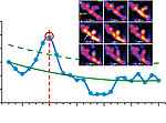
Capturing mitoflashes
[Galaxy workflows and tools]
[Paper]

Dorsal horn image registration
[Galaxy tool: landmark registration]
[Paper]


Selected peer-reviewed papers with methodological contributions
Gao Q, Rohr K. Non-rigid registration of time-lapse cell microscopy images improved by a semi-incremental optimization method. Proc. IEEE International Symposium on Biomedical Imaging (ISBI 2025), Houston, TX, USA, April 14-17, 2025, IEEE Piscataway, NJ. doi:10.1109/ISBI60581.2025.10981034 (IEEE ISBI 2025 Best Paper Award)
Gao Q, Chagin VO, Cardoso MC, et al. Quantifying newly appearing replication foci in cell nuclei based on 3D non-rigid registration. Proc. IEEE International Symposium on Biomedical Imaging (ISBI 2022), Kolkata, India, March 28-31, 2022, IEEE Piscataway, NJ. doi:10.1109/ISBI52829.2022.9761689
Wang D, Gao Q, Schaefer I, et al. TRPM3-mediated dynamic mitochondrial activity in nerve growth factor-induced latent sensitization of chronic low back pain. Pain. 2022;163(11):e1115-e1128. doi:10.1097/j.pain.0000000000002642
Liu S, Bonalume V, Gao Q, et al. Pre-synaptic GABAA in NaV1.8+ primary afferents is required for the development of punctate but not dynamic mechanical allodynia following CFA inflammation. Cells. 2022;11(15):2390. doi:10.3390/ cells11152390
Gao Q, Chagin VO, Cardoso MC, et al. Non-rigid registration of live cell nuclei using global optical flow with elasticity constraints. Proc. IEEE International Symposium on Biomedical Imaging (ISBI 2021), Nice, France, April 13-16, 2021, IEEE Piscataway, NJ, 1457-1460. doi:10.1109/ISBI48211.2021.9434064
Li R, Gao Q, Rohr K. Multi-object dynamic memory network for cell tracking in time-lapse microscopy images. Proc. IEEE International Symposium on Biomedical Imaging (ISBI 2021), Nice, France, April 13-16, 2021, IEEE Piscataway, NJ, 1029-1032. doi:10.1109/ISBI48211.2021.9433828
Gao Q, Rohr K. A global method for non-rigid registration of cell nuclei in live cell time-lapse images. IEEE Trans Med Imaging. 2019;38(10):2259-2270. doi:10.1109/TMI.2019.2901918
Adolf F, Rhiel M, Hessling B, Gao Q, et al. Proteomic Profiling of Mammalian COPII and COPI Vesicles. Cell Rep. 2019;26(1):250-265.e5. doi:10.1016/j.celrep.2018.12.041
Kontextspalte
Contact
For further information or to discuss your project, please contact:
Dr. Qi Gao
Computational Bioimage Scientist
Tridomus C410
Phone +49 621 383-71415
qi.gao@medma.uniheidelberg.de